

Understanding the complex anatomy of our bodies is key to maintaining physical wellness. In this anatomy lesson, we’re diving into the fascinating world of human anatomy, specifically exploring the anatomy of the adductor muscles. These key muscles, namely the adductor magnus, adductor longus, adductor brevis, pectineus, and gracilis, play a critical role in our daily movements and functionality of the lower body. We’ll explore the location, shape, size, origin, insertion, innervation, and blood supply of these muscles. We’ll also delve into their diverse range of functions and discuss strength training and stretching exercises designed specifically for them.
Functional anatomy of the adductor muscles
The adductor muscles of the leg make up the inner thigh. They are a cluster of five muscles that make up a relatively large muscle group when taken as a whole. The bulk of this muscle group starts where the leg meets the body and travels about halfway down the lower leg.

Muscles of the adductor group
Adductors consist of five muscles:
1. Adductor magnus
The adductor magnus is the largest and the strongest adductor muscle, as the name implies. It is also the deepest adductor, so many other adductors sit on top of it. In addition, it has the lowest insertion point of any adductor muscle that inserts on the femur.

2. Adductor longus
The adductor longus is, ironically, not as long as the adductor magnus, although it sits on top of the magnus. In fact, it is about half as long as the magnus. It is longer than the next muscle that we discuss (brevis), so that is where the name comes from. It is a relatively simple one-joint muscle.
3. Adductor brevis
The adductor brcvis is the shortest of the three main adductor muscles. The word brevis means “brevity” or “brief”, so the name makes sense. The adductor brevis sits somewhat superior and deep to the adductor longus, but it is superficial compared with the adductor magnus.

4. Pectineus
The pectineus muscle is the most superior adductor at its insertion point. It starts on the pubic bone, close to the ilium. Because of its anterior origin, it is a relatively strong hip flexor, working with the iliopsoas. It inserts in a similar place to the adductor brevis. The upper adductor muscles (pectineus, brevis, and longus) are often thought of as groin muscles, and when people say that they pulled a groin muscle, it is often one of these muscles.
5. Gracilis
The gracilis is the final adductor muscle. It is long and thin, about the width and thickness of a man’s belt. Gracilis runs from the pubic bone all the way down the inside of the leg to the tibia. It is the only two-joint adductor muscle. Because it inserts on the tibia, it can help flex the knee, but it is weak in that action. This muscle is the most medial of the adductors. If your thighs rub together when you walk, your gracilis muscles are touching.

Muscle location and shape
The adductor muscles fan out along the medial part of your thigh, which is the inner thigh area. They tend to look like a lump of muscle in the inner, upper thigh. Each of them has a unique shape and location within this collective. Most of the mass of the adductors is above the halfway point in the thigh.
The adductor longus, brevis, and magnus are the primary muscles that form the bulk of the adductors, and they spread across the inner thigh. The adductor longus sits right below the pubic bone and is somewhat elliptical in shape, easily recognized by its long, slender appearance. The adductor brevis, meanwhile, is a small, somewhat fan-shaped muscle found just beneath the adductor longus. Finally, the adductor magnus is the most substantial of the three. It spans the large area from the ischium to the back part of the femur and tibia, resembling the shape of a large triangle.
The pectineus, a flat quadrate muscle, overlaps the adductor brevis and longus near the pubic bone. On the other hand, the gracilis is the most superficial muscle in this group, being noticeably thin and flat, often referred to as a long strap muscle.
Points of origin and insertion
All the adductor muscles originate on the pubic bone, at least in part, and all but one (gracilis) insert on the femur.
| Adductor muscles | Muscle origin | Muscle insertion |
|---|---|---|
| Adductor magnus | Pubic bone (inferior ramus of the pubis); Ischium (ishiam tuberosity and ramus of the ischium) | Linea aspera of the ferum; Adductor tubercle of the femur |
| Adductor longus | Pubic bone | Linea aspera of the ferum |
| Adductor brevis | Pubic bone | Linea aspera of the ferum |
| Pectineus | Anterior pubic bone | Proximal femur between lesser trochanter and linea apera |
| Gracialis | Pubic bone | Medial procimal tibia |
A memory trick to remember the insertion points is to think of “peanut butter loves mashed grapes” (mashed grapes are jelly). Going from top to bottom, the insertion of the muscles are pectineus, brevis, longus, magnus, and gracilis.
As you reflect on this information, it’s clear to see how intimately connected the anatomy of the adductor muscles is to your movement. Appreciating their points of origin and insertion allows us to truly grasp the intricate puzzle that is our muscular system.
Primary functions of the adductor group
As the name implies, they all perform adduction of the hip, both horizontal and vertical.
| Adductor muscles | Functions |
|---|---|
| Adductor magnus | Overall - adduction of the hip; Anterior part - assist in hip flexion; Posterior part - assist in hip extension |
| Adductor longus | Adduction of the hip, helps with hip flexion |
| Adductor brevis | Adduction, helps with hip flexion |
| Pectineus | Adduction and flexion of the hip |
| Gracialis | Adduction of the hip; Assist with knee flexion (very weak) |
Size of the adductor muscles
Generally, the size of the adductor muscles is smaller compared to the more substantial, more dominant muscles of the leg, like the quadriceps femoris and the hamstrings. However, don’t let the size fool you! The adductors pack a considerable punch in functionality. They are vital for stability, mobility, and providing balance during a wide range of activities, proving that it’s not always their size that determines their value.
We can visualize this size difference by looking at their respective locations. The quadriceps femoris, located at the front of the thigh, is particularly large and robust, making up a significant portion of the thigh mass. The hamstrings, found at the back of the thigh, also have considerable bulk. In comparison, the adductor muscles, situated in the inner thigh, appear somewhat smaller. Nonetheless, what they may lack in size, they make up for in their vital role in bridging the actions of the quadriceps and hamstrings.
Innervation & blood supply
The innervation and blood supply of the adductor muscles is a crucial component of their function. These elements support muscle activity, by providing the sensory and motor responses necessary for functioning, in addition to delivering oxygen and nutrients to the muscles.
Primarily, the adductor muscles are innovated by the obturator nerve (L2-L4). However, some exceptions do exist. Specifically, the adductor magnus is innervated by both the obturator and sciatic nerves.
In terms of blood supply, branches of the femoral and obturator arteries typically provide the blood to the hip adductors.
Adductors vs Abductors
The opposite muscles to the adductors are abductors. It’s all in the names. Adductors “add” the limb to the center of the body, while abductors “abduct” or take away the limb from the center.
The adductors are a group of muscles located in your inner thigh that serve to draw your leg toward the midline of your body.
On the flip side, your abductor muscles are located on the outer side of your thighs and hips. When these muscles work, they move your legs away from your body’s midline. This is a key function in actions like kicking a ball sideways or stepping to the side. The primary abductor muscles include the gluteus medius, gluteus minimus, and tensor fasciae latae.
Best bodybuilding exercises to grow your adductors
Training the adductor muscles in the most effective way involves a combination of isolation and compound exercises. You can emphasize the adductors in the gym by using a wide stance on most compound bilateral exercises. Exercises like squats, lunges, and leg press with a wider stance can engage multiple muscle groups, including the adductors. While these exercises don’t isolate the adductors, they contribute to overall leg and core strength, and flexibility—factors crucial to adductor health and performance. Going extra low also emphasizes the adductors. Using single-leg variations works both the adductors and abductors more.
However, in order to really hit your adductors you will have to use exercises machines and isolation exercises. These directly target the adductor muscles. It is totally impossible to isolate the adductors using free weights alone. Popular exercises include the seated adductor machine and standing cable hip adductions. By focusing on muscle contractions and slow, controlled movements, you can exert greater force on the adductors and stimulate growth.
Here’s the list of the most important exercises for the adductors (ranked in order of importance):
- Adductor machine (machine adductions);
- Standing machine hip adductions;
- Cable hip adductions;
- Sumo deadlifts;
- Wide-stance squats and leg presses;
- Medicine ball leg raise (ball held between feet);
 Low pulley cable hip adduction
 Cable Hip Adduction
 Standing Machine Hip Adductions
 machine hip adductions
 wide-stance squats
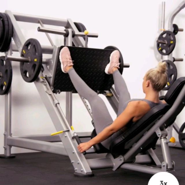 wide-stance leg presses
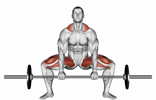 sumo deadlifts
Stretching & flexibility exercises for the adductors
- Groin Stretch: Stand with your legs wide apart. Shift your weight to one side, bending the knee and reaching your groin area toward the floor. Hold for 20-30 seconds, then switch sides. This stretching exercise aims to improve the flexibility of your adductors.
- Baddha Konasana or Butterfly Pose: Sit on the ground and bring the soles of your feet together. Use your elbows to push your knees toward the floor. Hold for 20-30 seconds, rest, and repeat. This yoga pose stretches and opens up your adductor muscles.
- Side lunges: Extend one leg to the side while bending the other knee, as if you were going to sit back into a chair. This exercise stretches the inner thigh and adductor muscles.
- Lying Hip Adductor Stretch: Lay down on your back and let both your knees fall outward with your feet touching. This position should stretch the adductor muscles in your inner thigh and groin area.
- Seated Forward Fold: While seated with your legs extended out in front of you, reach for your toes while keeping your back straight. This also helps in stretching your hamstrings along with your adductors if you spread your legs apart.
Common issues & injuries associated with adductors
The adductor muscles, while relatively resilient, are not exempt from the risk of injuries or strains. They play an active role in our everyday movements, constantly called upon for actions such as walking, running, and jumping. As a result, these muscles can be subject to injuries, often due to disproportionate strain or unusual movement patterns.
- Adductor muscle strains or ‘groin strains’ are common occurrences, particularly amongst athletes who often engage in strenuous activities involving heavy lifting, repetitive motions, or sudden direction changes. This generally manifests as a sharp pain in the inner thigh, often accompanied by swelling and bruising. This injury can happen when the adductor muscles are stretched beyond their limit, or experience sudden contractions.
- Muscle imbalances can also be problematic for the adductors. In particular, a malposition of the sacroiliac joint can restrict the function of the hip adductors, causing pain, discomfort, and potentially persistent complications if left unaddressed.
Of course, it’s always the best protocol to prevent these injuries. Regular strength and flexibility training aimed at your adductor muscles can often keep them in top shape, minimizing the risk of injury. But if you do notice any discomfort or persisting pain, seeking medical advice should be an immediate step to prevent further aggravation.
The group commonly known as the adductors consists of five medial muscles that move the thigh toward the midline of the body. The gracilis muscle is the most superficial, the pectineus is the most proximal, and the adductor longus is immediately lateral to the gracilis. The adductor brevis is located behind the adductor longus. The adductor magnus, the largest of the group, is on the medial side of the thigh. All five adductor muscles originate from different locations on the os pubis (pubic bone) and are inserted into the femur. The gracilis is the only one that inserts into the tibia. All five muscles assist with adduction, flexion, and rotation of the thigh.
