


Every time you perform a lateral raise, squat, bench press, or triceps extension, tendons, muscles, skin, and bones move relative to one another. In several high-friction areas, tiny fluid-containing structures called bursae help those tissues glide more smoothly against each other.
Quick Answer
A bursa is a small, thin sac located between tissues that move against one another, commonly near synovial joints. Most anatomical bursae are lined by synovial membrane and contain a small amount of lubricating fluid. Their primary function is to reduce friction and facilitate smooth movement between structures such as tendons, muscles, skin, and bone. The singular term is bursa; the plural is bursae. Major joints such as the shoulder, hip, and knee contain multiple bursae positioned where mechanical friction or pressure commonly occurs.
1. Where Are Bursae Found?
It’s worth correcting a common assumption right away: bursae aren’t simply “inside joints.” Most anatomical bursae sit around joints, interposed between structures that move against one another — one of the accessory structures covered in our joints in the human body guide — not floating freely within the synovial joint cavity itself, though some do communicate with that cavity.
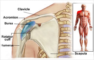
Bursae are found near most major joints in the body, particularly where friction or pressure commonly occurs — the shoulder, elbow, hip, knee, and ankle/heel region among the most relevant to lifters. The adult body contains roughly 160 bursae in total, though only a relatively small number are regularly relevant to resistance training.
2. What Is a Bursa Made Of?
A typical synovial bursa is a thin connective-tissue sac, lined internally by a synovial membrane, containing a small amount of lubricating synovial fluid — the same fluid covered in depth in our dedicated synovial fluid guide.
Here’s a nuance worth knowing: a healthy bursa isn’t a large, visibly swollen “water balloon.” Imaging literature notes that bursae are often most clearly visualized specifically when they’re distended with fluid — meaning a normal, non-irritated bursa is frequently a thin, low-profile structure that doesn’t stand out much on imaging at all. A healthy bursa is normally a thin, low-friction interface rather than a visibly swollen fluid-filled pouch.
3. What Does a Bursa Do?
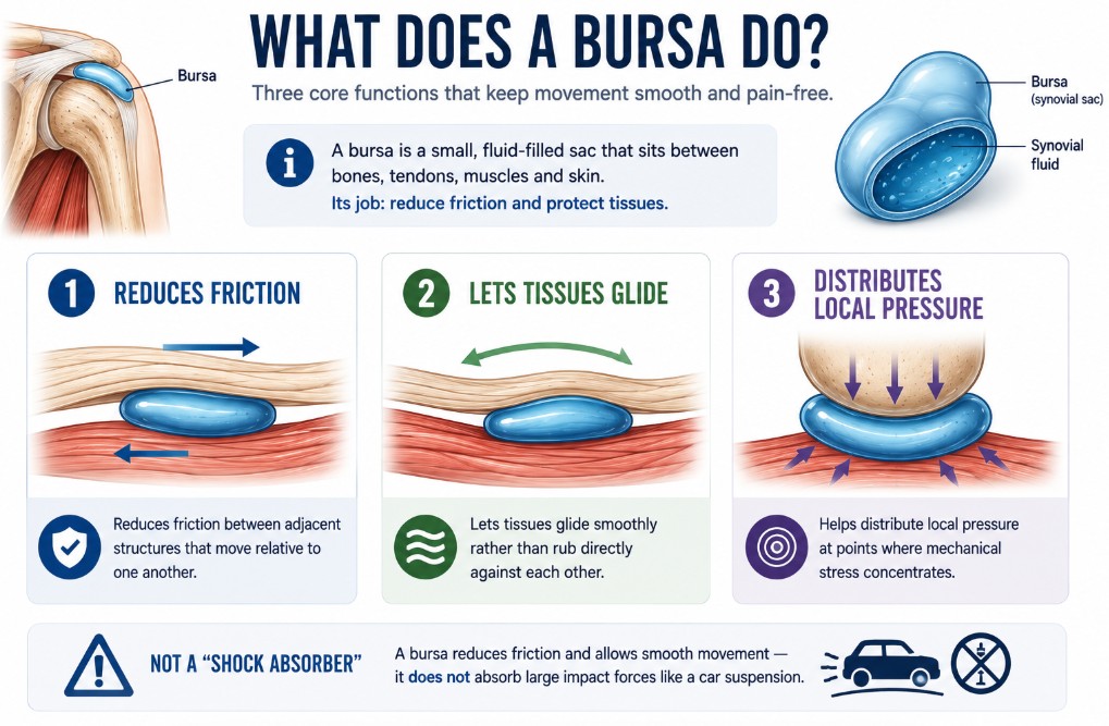
Three core functions:
- Reduces friction between adjacent structures that move relative to one another.
- Lets tissues glide smoothly rather than rub directly against each other.
- Helps distribute local pressure at points where mechanical stress concentrates.
Be careful with the word “shock absorber” here, since it tends to suggest something closer to automotive suspension—a bursa’s primary role is reducing friction and facilitating smooth movement, not absorbing large impact forces the way that phrase implies.
4. How Bursae Reduce Friction During Movement
Picture a tendon repeatedly moving over a bony prominence during a lift. Without a low-friction interface at that point, the tendon would experience much more direct tissue-on-tissue friction with every repetition. A bursa sits between these structures, allowing relative sliding with minimal resistance.
A bursa does not generate movement; it makes movement between neighboring structures mechanically smoother. That distinction matters — a bursa is a passive mechanical interface, not an active contributor to force production the way muscle is.
5. Types of Bursae
Classification here is fairly consistent across anatomy sources, organized by the specific tissues a given bursa sits between (Mercadante & Marappa-Ganeshan, 2022):
- Subcutaneous bursae — between skin and bone. Examples include the prepatellar bursa (front of the knee) and olecranon bursa (tip of the elbow).
- Subtendinous bursae — between tendon and bone.
- Submuscular bursae — between muscle and bone, or between adjacent muscles.
- Subfascial bursae — between fascia and deeper structures.
6. Constant vs Adventitious Bursae
This is a genuinely interesting distinction that most basic anatomy content skips entirely.
Constant (anatomical) bursae develop during embryonic development at predictable, consistent locations — the same bursae, in the same places, in essentially everyone. Adventitious bursae, by contrast, aren’t present at birth and can develop later in life at sites of repeated friction or pressure that weren’t originally bursa locations at all (Mercadante & Marappa-Ganeshan, 2022).
In other words, the body isn’t working from a completely fixed anatomical map here — it can form new bursae in response to a chronic mechanical environment, a small but genuinely interesting example of tissue responding to the demands placed on it over time.
7. Major Bursae Lifters Should Know
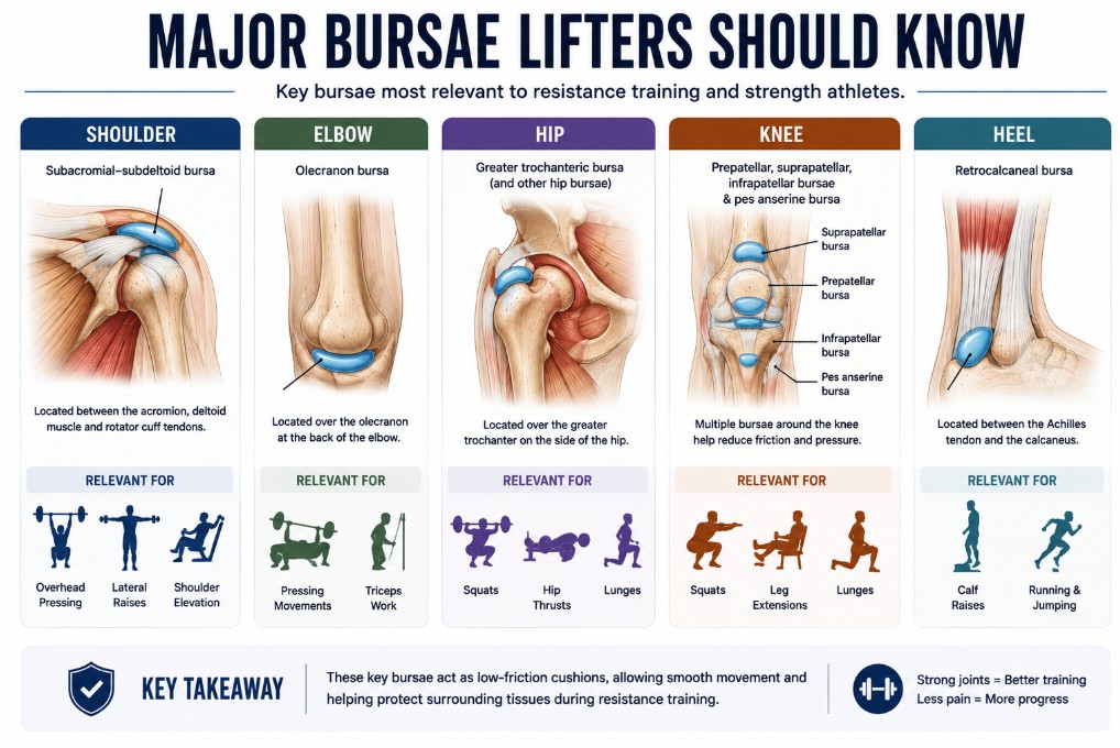
Cataloging all ~160 bursae in the body wouldn’t be useful here — this guide focuses on the ones most relevant to resistance training.
- Shoulder: the subacromial-subdeltoid bursa, relevant to overhead pressing, lateral raises, and other movements involving shoulder elevation — covered in more anatomical depth in our shoulder joint anatomy guide.
- Elbow: the olecranon bursa, at the back of the elbow.
- Hip: the greater trochanteric bursa and other regional hip bursae — see our hip joint anatomy guide for the surrounding structures.
- Knee: several bursae including the prepatellar, suprapatellar, and infrapatellar bursae, along with the pes anserine region — covered further in our knee joint anatomy guide.
- Heel: the retrocalcaneal bursa, situated between the Achilles tendon region and the calcaneus.
8. Bursa vs Synovial Fluid: What’s the Difference?
Worth stating plainly, especially given how often these terms get blurred together: a bursa is a structure; synovial fluid is a substance. A bursa is the sac itself — the anatomical structure. Synovial fluid is the lubricating liquid contained within it (and within synovial joints more broadly, as covered in our dedicated synovial fluid guide). One is a container; the other is what’s inside it.
9. Bursa vs Tendon Sheath
A related structure worth briefly distinguishing: the tendon sheath, a bursa-like structure that wraps around a tendon along part of its course, rather than sitting between two separate structures the way a typical bursa does.
The distinction in short: a bursa sits between adjacent structures that move relative to one another. A tendon sheath wraps around a single tendon, providing a low-friction tunnel as that tendon glides through it — different geometry serving a related friction-reducing purpose.
10. What Is Bursitis?
Worth a brief, properly scoped overview here, since it’s a major part of what people search for around this topic — though this guide stays at the overview level rather than becoming a diagnostic or treatment resource.
Bursitis refers to inflammation or pathological irritation of a bursa, often associated with increased fluid volume and changes to the synovial lining (Williams, Jamal & Sternard, 2023). It can arise in several different contexts: repetitive pressure, friction, direct trauma, underlying inflammatory conditions, and in some cases infection. This guide won’t cover diagnosis, treatment, or rehabilitation protocols — that’s the domain of dedicated injury-focused content, not a general anatomy guide.
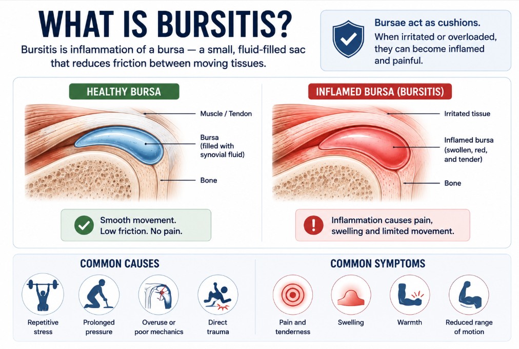
11. Why Do Lifters Get Irritated Bursae?
Rather than a blanket claim that “lifting causes bursitis,” it’s more accurate to name the range of potential contributors: repeated compression at a specific site, abrupt changes in training workload, repetitive positioning that concentrates stress in one location, direct pressure (kneeling on a hard surface, for instance), individual anatomy, previous injury history, and technique or exercise selection within that broader context.
Pain near a bursa does not prove that the bursa is the source of that pain. Multiple structures often sit in close proximity—tendon, joint capsule, adjacent bursae, and more—and identifying a specific bursa as the actual cause of discomfort requires more than noting where the pain happens to be.
12. Does Heavy Lifting Damage Bursae?
Not automatically — and it’s worth being direct about that rather than hedging indefinitely.
Bursae exist precisely in the mechanical environments where tissues repeatedly move against each other under load. The presence of friction, movement, and mechanical demand at a bursa isn’t itself the problem — bursae are built for exactly that environment. A genuine issue arises when load or pressure at a specific site exceeds what the tissue can currently tolerate, or when an underlying pathological process is already present. That’s a meaningfully different claim than “heavy lifting damages bursae” as a general rule, and this guide won’t collapse that distinction for the sake of a simpler-sounding answer.
13. Can You Strengthen a Bursa?
Not in the same sense you’d strengthen a muscle — and this is worth stating clearly, because the framing itself is usually the wrong one.
A bursa isn’t contractile tissue, and it doesn’t hypertrophy or adapt to training the way muscle does. You do not train a bursa directly. The practical goal is to build the capacity of the surrounding tissues and movement patterns, rather than trying to “strengthen” the bursa itself. Training the muscles, tendons, and joint structures around a given bursa — and progressively building tolerance for the movements and positions that load that area — is the relevant training target, not the bursa in isolation.
14. Training Implications for Lifters

Five practical principles, drawn from everything above.
- Don’t fear normal friction and joint movement. Bursae exist specifically to make that kind of movement mechanically smoother, not to be protected from it entirely.
- Progress repetitive loading gradually, particularly in movements that concentrate repeated stress at a specific site.
- Avoid unnecessary direct pressure on irritated superficial bursae — a practical, common-sense consideration, not a treatment prescription.
- Exercise selection isn’t universally “bursa-friendly” or “bursa-damaging.” Individual anatomy, technique, and training context all matter more than a fixed list of good or bad exercises.
- Persistent swelling or loss of function deserves proper assessment, not pushing through or self-diagnosing.
Coach’s Note: Don’t think of bursae as fragile water balloons you need to protect from movement. They are normal mechanical interfaces positioned where tissues repeatedly slide against one another. For lifters, the goal is not to avoid loading but to build enough capacity for the movements and training volumes you perform.
Key Takeaways
- ✓A bursa is a small fluid-containing sac that reduces friction between moving tissues — the plural is bursae.
- ✓Bursae are commonly located near synovial joints but are not simply “inside” the joint itself.
- ✓They can sit between tendon and bone, muscle and bone, skin and bone, or fascia and deeper structures.
- ✓Some bursae are constant and present from embryonic development; others are adventitious, developing later at sites of chronic friction or pressure.
- ✓A bursa is a structure; synovial fluid is the substance found inside it and inside synovial joints more broadly.
- ✓Bursitis refers to pathological inflammation or irritation of a bursa — not a diagnosis this guide covers in depth.
- ✓Normal resistance training does not automatically damage bursae — they exist specifically for mechanically active environments.
- ✓Lifters don’t need to “strengthen” bursae directly — the practical target is the surrounding muscle, tendon, and joint capacity.
Frequently Asked Questions
What is a bursa? +
A bursa is a small, thin sac located between tissues that move against one another, commonly near synovial joints. Most are lined by synovial membrane and contain a small amount of lubricating fluid, and their primary role is reducing friction between structures like tendon, muscle, skin, and bone.
What is the function of a bursa? +
A bursa reduces friction, allows tissues to glide smoothly against one another, and helps distribute local pressure at points where mechanical stress concentrates during movement.
Where are bursae located? +
Bursae are found around most major joints in the body — commonly the shoulder, elbow, hip, knee, and heel/ankle region — positioned between structures such as tendon and bone, muscle and bone, or skin and bone, wherever friction or pressure commonly occurs.
How many bursae are in the human body? +
The adult body contains roughly 160 bursae, though only a relatively small subset are regularly relevant to resistance training and everyday movement.
What is inside a bursa? +
A bursa contains a small amount of synovial fluid, lubricating the interior surface lined by synovial membrane. A healthy bursa typically contains only a thin film of fluid, not a large visible pool.
What is the difference between a bursa and synovial fluid? +
A bursa is a structure — the sac itself. Synovial fluid is a substance — the lubricating liquid found inside that sac (and inside synovial joints more broadly). One is a container; the other is what’s inside it.
What is bursitis? +
Bursitis is inflammation or pathological irritation of a bursa, often involving increased fluid and changes to the synovial lining. It can result from repetitive pressure, friction, direct trauma, inflammatory conditions, or occasionally infection.
Can exercise irritate a bursa? +
It can, particularly with repeated compression, abrupt workload changes, repetitive positioning, or direct pressure at a specific site. But this isn’t automatic — bursae exist specifically to function in mechanically active environments, and irritation typically relates to load exceeding current tissue tolerance rather than movement itself being the problem.
Does weightlifting cause bursitis? +
Not automatically. Weightlifting places bursae in the mechanical environment they’re built to handle. Bursitis is more closely associated with load exceeding a tissue’s current tolerance, sudden workload spikes, direct repeated pressure, or pre-existing pathology, rather than resistance training itself being an inherent cause.
Can you strengthen a bursa? +
Not in the way you’d strengthen a muscle. A bursa isn’t contractile tissue and doesn’t adapt to training directly. The practical training target is building the capacity of the surrounding muscles, tendons, and joint structures, along with progressively increasing tolerance for the movements that load that area.
References
- Mercadante, J. R., & Marappa-Ganeshan, R. (2022). Anatomy, Skin Bursa. StatPearls. StatPearls Publishing. ncbi.nlm.nih.gov/books/NBK554438
- Williams, C. H., Jamal, Z., & Sternard, B. T. (2023). Bursitis. StatPearls. StatPearls Publishing. ncbi.nlm.nih.gov/books/NBK513340
