
Every press, row, raise, and pull-up asks your shoulder to do something almost no other joint in your body is asked to do: move through an enormous range of motion while staying stable enough to transmit real force. That combination — huge mobility, modest built-in stability — is not incidental. It’s the direct result of how the shoulder is constructed, and understanding that construction explains a lot about why shoulders behave the way they do under a barbell.
This guide builds on the general joint principles covered in our joints in the human body guide and applies them specifically to the shoulder — working from bone and cartilage, through the soft tissue that provides most of this joint’s stability, into the muscles that control it, and finally into what all of this means for how you train.
Quick Answer
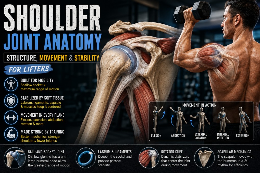
The shoulder joint most people mean when they say “shoulder” is the glenohumeral joint — a ball-and-socket joint between the head of the humerus and the shallow glenoid fossa of the scapula. Its socket covers roughly a quarter to a third of the humeral head, giving the shoulder the largest range of motion of any joint in the body. That same shallow fit means the shoulder has relatively little passive bony stability, so it relies heavily on the labrum, joint capsule, ligaments, and — most importantly for lifters — the rotator cuff and surrounding musculature to stay properly positioned under load.
1. The Shoulder Isn’t One Joint — It’s a Complex
When lifters talk about “shoulder pain” or “shoulder mobility,” they’re almost always talking about one specific joint — the glenohumeral joint. But the functional shoulder is actually a system of four joints working together:

- Glenohumeral joint — the ball-and-socket joint between humerus and scapula; the main subject of this guide
- Acromioclavicular (AC) joint — where the clavicle meets the acromion of the scapula
- Sternoclavicular (SC) joint — where the clavicle meets the sternum, the only bony connection between the arm and the axial skeleton
- Scapulothoracic joint — not a true anatomical joint, but the functional gliding interface between the scapula and the rib cage, essential to how the whole shoulder complex moves
This guide focuses primarily on the glenohumeral joint, since that’s what “shoulder joint anatomy” means to almost everyone searching for it — but the other three keep showing up throughout this guide, because the glenohumeral joint literally cannot function properly in isolation from them.
2. Bones of the Shoulder Complex

Three bones form the shoulder complex: the humerus (upper arm bone, contributing its rounded head), the scapula (shoulder blade, contributing the glenoid fossa, acromion, and coracoid process), and the clavicle (collarbone, linking the shoulder to the sternum).
This guide won’t re-cover general skeletal anatomy here — for the fuller picture of these bones in context, see our interactive human skeleton tool and major bones of the skeletal system guide. What matters for this guide is how two of these bones — the humerus and scapula — meet to form the glenohumeral joint, which is where the real anatomical story begins.
3. The Glenohumeral Joint: A Ball-and-Socket Built for Mobility
The glenohumeral joint pairs the head of the humerus — large, round, and covered in articular cartilage — with the glenoid fossa, a shallow, pear-shaped depression on the scapula, also cartilage-covered where the two bones meet.
The mismatch between these two surfaces is the single most important anatomical fact about the shoulder. The glenoid fossa covers only about a quarter to a third of the humeral head, and comparisons of the surface area between the two structures put the ratio at roughly 4:1 in the humeral head’s favor (StatPearls, 2025). Put simply: the socket is nowhere near big enough to fully contain the ball.
This is deliberate, in an evolutionary sense — that shallow fit is exactly what allows the humeral head to rotate and translate through an enormous range of motion without the bone geometry itself blocking the movement. But congruency (how well the two joint surfaces match each other) isn’t just about overall size. Both surfaces are covered in articular cartilage, distributing load and reducing friction as the humeral head glides and rotates across the glenoid, and even this cartilage layer is not uniform — cartilage thickness varies across the glenoid surface, generally thinner centrally where contact is greatest and thicker toward the periphery (Cuéllar, Ruiz-Ibán & Cuéllar, 2017). This isn’t a flaw in the design — it’s a joint built to prioritize freedom of movement over a tight mechanical fit, which sets up everything else in this guide.
4. The Glenoid Labrum
Because the bony socket alone provides so little containment, the shoulder relies on a specialized structure to improve the fit: the glenoid labrum, a ring of fibrocartilage attached around the rim of the glenoid fossa.
The labrum does several jobs at once. It deepens the socket — one anatomical study measuring this effect found the labrum increases the effective length of the glenoid surface by roughly a third (Almajed et al., 2022) — and it also serves as the attachment point for the glenohumeral ligaments and the long head of the biceps tendon, tying several stabilizing structures together at a single anatomical hub. Beyond simply deepening the socket, the labrum contributes to a suction/negative-pressure effect within the joint and helps maintain consistent contact between the humeral head and glenoid cartilage.
Its practical importance becomes clearest when it’s damaged. Cadaveric research removing the anteroinferior portion of the labrum found the contact area between the humeral head and glenoid dropped by roughly 7–15%, while contact pressure in that same region rose by 8–20% (Almajed et al., 2022) — a meaningful shift in how load gets distributed across the joint surface from a structure most lifters never think about until something goes wrong with it.
Why This Structure Matters Under Load
A labrum that’s doing its job properly means load gets distributed across a reasonably large, well-contained contact area. A compromised labrum concentrates more of that same load onto a smaller area of cartilage — which is part of why labral pathology (a topic for a dedicated injury-focused guide, not this one) tends to matter mechanically, not just as a source of pain.
5. The Joint Capsule and Glenohumeral Ligaments
Surrounding the entire glenohumeral joint is the joint capsule — a fibrous sleeve running from the rim of the glenoid to the anatomical neck of the humerus. Compared to the capsules of more constrained joints, the glenohumeral capsule is relatively loose and capacious, particularly along its inferior aspect — a design that permits the joint’s extreme range of motion rather than restricting it the way a tighter capsule would at a more congruent joint.
Within the capsule, three regional thickenings form the glenohumeral ligaments — passive stabilizing structures that don’t act uniformly throughout the joint’s range, but instead tighten and slacken depending on arm position:
| Ligament | Generally Taut When | Primary Restraint |
|---|---|---|
| Superior (SGHL) | Arm adducted at the side | Inferior humeral head translation in the dependent arm |
| Middle (MGHL) | Mid-range abduction with external rotation | Anterior-posterior translation in the mid-range |
| Inferior (IGHL) complex | Abduction combined with external or internal rotation | The primary restraint against anterior shoulder dislocation |
The inferior glenohumeral ligament complex is generally regarded as the most clinically significant of the three, since it becomes the primary passive restraint in the abducted, externally rotated position — a position that shows up directly in movements like the bottom of a bench press or the loaded top of a lateral raise, which this guide returns to later (Gasbarro et al., 2017).
Worth being precise about what these ligaments are and aren’t doing: they don’t function like a static seatbelt that only engages at a single end-range moment. Each ligament’s contribution shifts continuously as the joint position changes, providing restraint across a spectrum of arm positions rather than one fixed “locked” position.
6. Bursae of the Shoulder
A handful of fluid-filled sacs called bursae reduce friction between tissues that would otherwise rub against each other during shoulder movement. The most clinically relevant is the subacromial (subdeltoid) bursa, sitting between the rotator cuff tendons below and the acromion and deltoid above — a location that puts it directly in the path of the space rotator cuff tendons need to glide through during overhead movement.
This guide won’t go deep into bursal pathology here — bursitis and its relationship to shoulder mechanics deserve their own dedicated treatment, which is planned as part of this connective tissue series. For now, the anatomical point that matters is simply that this bursa exists precisely because the rotator cuff tendons and the bony arch above them are close enough that friction-reduction becomes necessary during normal shoulder function — a spatial relationship that becomes relevant again later in this guide.
7. Muscles and Tendons: The Rotator Cuff and Beyond
Given how little passive bony stability the shoulder has to begin with, the muscles crossing this joint aren’t optional extras — they’re doing a large share of the work that a deeper socket would otherwise handle on its own.

The rotator cuff
Four muscles make up the rotator cuff — supraspinatus, infraspinatus, teres minor, and subscapularis. Their combined job goes well beyond producing rotation: they work together as coordinated force couples, compressing the humeral head into the glenoid fossa (a mechanism called concavity compression) to keep it centered throughout the joint’s enormous range of motion. The subscapularis anteriorly and the infraspinatus/teres minor posteriorly form one such force couple, balancing anterior-posterior forces on the humeral head, while the rotator cuff as a whole counteracts the superior pull of the deltoid during arm elevation, preventing the humeral head from migrating upward into the subacromial space (Akhtar, Richards & Monga, 2021).

The deltoid
The deltoid — anterior, middle, and posterior heads — is the shoulder’s primary prime mover, generating most of the raw force behind abduction and flexion. But the deltoid’s line of pull, acting alone, would tend to translate the humeral head upward rather than rotate it cleanly within the socket. This is precisely why the deltoid and rotator cuff function as a paired system rather than independently: the cuff’s compressive, centering action allows the deltoid’s larger force to produce clean rotation instead of instability.
The long head of the biceps tendon
Often overlooked, the long head of the biceps tendon (LHBT) originates from the supraglenoid tubercle and superior labrum and runs an unusual intra-articular course through the joint before exiting into the bicipital groove. This anatomical path lets it function as a dynamic stabilizer in its own right — cadaveric research loading the tendon found it reduced anterior humeral head translation by roughly 43% and inferior translation by roughly 73% compared to an unloaded tendon (Alexander et al., 2013). This is worth knowing for one specific reason: the LHBT’s attachment directly onto the superior labrum is the anatomical link that connects labral health to biceps tendon health, a relationship this guide won’t go further into here, but one worth being aware of.
Scapular stabilizers
None of the above works properly if the scapula itself isn’t stable. Three muscle groups do most of that job: the serratus anterior (middle and lower fibers driving scapular upward rotation, posterior tilt, and external rotation), the trapezius (upper fibers elevating and retracting the clavicle, middle fibers acting as a medial stabilizer, lower fibers assisting both stabilization and upward rotation), and the rhomboids, which retract the scapula and help counter the lateral pull the serratus anterior places on it (Phadke, Camargo & Ludewig, 2009).
This is the missing piece in a lot of simplified shoulder content: rotator cuff strength alone doesn’t guarantee a well-functioning shoulder if the scapula it’s attached to isn’t being properly positioned and controlled in the first place.
8. Shoulder Movements: Glenohumeral and Scapular
It’s worth separating these into two distinct categories, because they happen at different joints and are produced by different muscles — even though, in real movement, they virtually always happen together.

Glenohumeral movements
- Flexion/extension — raising the arm forward, lowering it back
- Abduction/adduction — raising the arm out to the side, returning it to the body
- Internal/external rotation — rotating the humerus around its long axis, as in the top and bottom of an overhead press
- Horizontal abduction/adduction — moving the arm across the body versus away from it at 90° of abduction, as in a chest fly versus a reverse fly
Scapular movements
- Elevation/depression — shrugging the scapula up, or pulling it down
- Protraction/retraction — the scapula sliding forward around the rib cage, or pulling back toward the spine, as in the eccentric versus concentric phases of a bench press
- Upward / downward rotation — the scapula rotating so the glenoid fossa tilts upward (during overhead reaching) or returns to neutral
Separating these two categories isn’t just a naming convention. It sets up the next section directly, because neither category happens in isolation — they’re mechanically coupled.
9. Scapulohumeral Rhythm
Scapulohumeral rhythm describes the coordinated relationship between glenohumeral motion and scapulothoracic motion during arm elevation. As the arm goes overhead, the humerus doesn’t do all the work alone — the scapula rotates upward to keep the glenoid fossa properly oriented under the moving humeral head, maintaining a stable relationship between the ball and socket throughout the movement rather than letting the humeral head run out of usable socket partway through the lift.
The classic teaching model describes this as a fixed 2:1 ratio — roughly 2° of glenohumeral motion for every 1° of scapular upward rotation. It’s a useful conceptual starting point, but treating it as a fixed law doesn’t hold up against the data. Research directly measuring this relationship has found the ratio varies considerably across the arc of elevation and, notably for lifters, changes substantially with external load: one study found the ratio shifted from roughly 2.1:1 unloaded to as high as 4.5:1 under heavy resistance, with the scapula contributing proportionally more to the movement as load increased (McQuade & Smidt, 1998). Other work measuring the ratio across the full elevation arc found values ranging as widely as 0.9:1 to 40:1 depending on exactly where in the range of motion the measurement was taken, with an overall average closer to 2.3:1 (Scibek & Carcia, 2012).
The practical takeaway isn’t a specific number to memorize — it’s that scapulohumeral rhythm is not a fixed mechanical constant. It shifts with load, phase of the movement, and individual factors, which is exactly why scapular control isn’t a “nice to have” add-on for overhead lifting — it’s a variable that measurably matters more, not less, as the weight on the bar increases.

10. How the Shoulder Is Loaded During Major Exercises
Rather than treating every shoulder exercise as a single, undifferentiated “shoulder stress,” it’s more useful to look at what specific factors change the loading picture — joint position, moment arms, and the muscular and scapular demands that follow from them. Four common exercises illustrate this well.
Bench press. Grip width, shoulder abduction angle, and scapular position all independently change how load is distributed across the glenohumeral and acromioclavicular joints. A detailed musculoskeletal modeling study found that retracting the scapula and using a moderate rather than very wide grip reduced posterior shear forces at the glenohumeral joint and lowered rotator cuff activity, while wider grip widths increased acromioclavicular compression forces (Noteboom et al., 2024). In plain terms: bench press “shoulder stress” isn’t one fixed quantity — it changes meaningfully with setup choices well within a lifter’s control.
Overhead press. This movement asks the shoulder to combine elevation with scapular upward rotation while the joint moves through progressively larger portions of its available range — precisely the loading condition where the scapulohumeral rhythm research covered above becomes directly relevant. As covered earlier, scapular contribution to the movement increases as external load increases, meaning heavier overhead pressing places proportionally greater demand on scapular control, not just on the deltoid and rotator cuff.
Lateral raise. As the arm moves into abduction, the moment arm between the resistance and the shoulder joint lengthens considerably, increasing the torque demand on the muscles producing and controlling that movement — the same moment-arm principle covered in our joints in the human body guide, applied specifically here to the lateral raise. This is part of why relatively light loads can still feel demanding in this position, and why strict form (versus using momentum to swing the arm up) meaningfully changes the loading profile.
Pull-up/row. These movements combine humeral extension or adduction with scapular retraction and depression, placing different demands on the shoulder stabilizers than pressing movements do. A pull-up and a bent-over row both require this coordination, though from different starting positions. Because the glenohumeral and scapular contributions are both substantial and coordinated, poor scapular control during pulling movements (letting the scapula elevate excessively, for instance) can shift load onto structures — like the levator scapulae or upper trapezius — that weren’t intended to be the primary stabilizers for that movement.
11. Individual Anatomical Variation
Not every shoulder is built to identical specifications, and some of that variation meaningfully changes what’s “normal” for a given lifter. Glenoid orientation varies between individuals — average retroversion (the glenoid facing slightly backward relative to the scapula) is commonly cited around 7°, while the humeral head itself typically shows considerably more retroversion, on the order of 30° relative to the shaft (Cuéllar, Ruiz-Ibán & Cuéllar, 2017). Capsular laxity also varies — some individuals have naturally more capacious, compliant capsules (sometimes associated with broader joint hypermobility), which shifts how much a given shoulder relies on active muscular control versus passive restraint to stay stable under load.
None of this variation is inherently good or bad. It simply means two lifters with textbook-normal shoulders can have meaningfully different available range of motion, different natural end-feels, and different tolerance for specific positions — which is exactly why blanket ROM prescriptions (“everyone needs to bench press to this exact depth”) don’t hold up well against real anatomical variability.
12. Common Shoulder Problems in Lifters — Overview
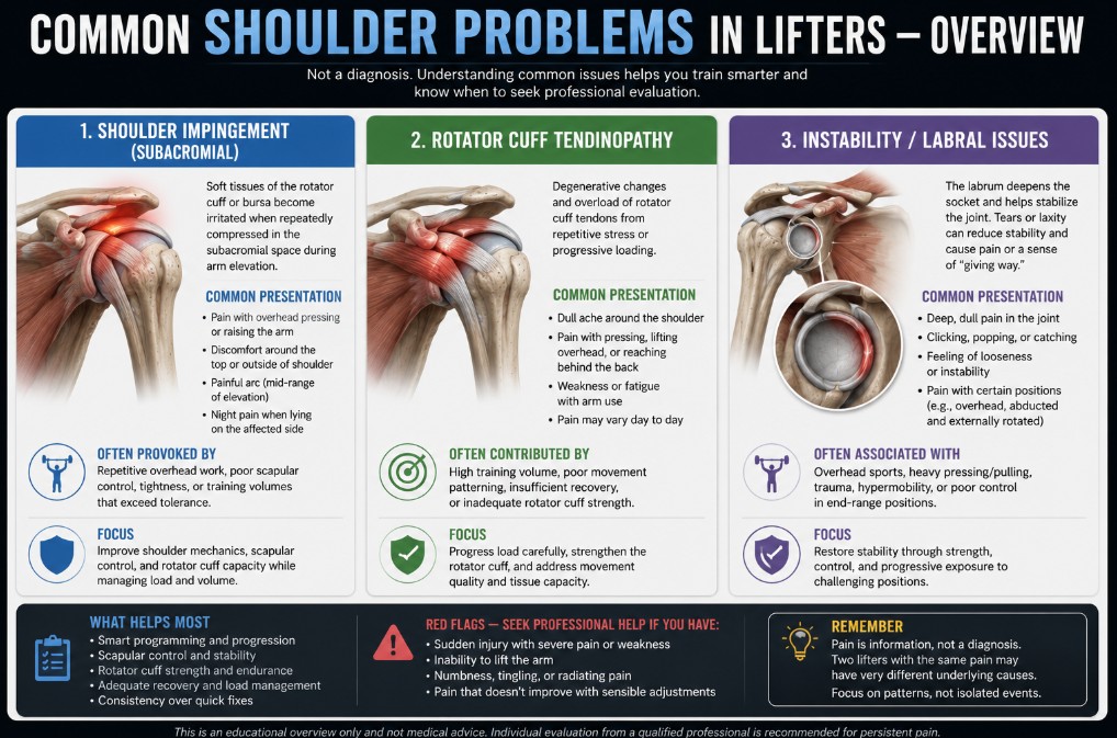
This guide has stayed focused on healthy anatomy and biomechanics throughout, and that continues here — this is a map, not a diagnostic or treatment guide.
Lifters most commonly encounter subacromial impingement (pain associated with compression of structures in the subacromial space), rotator cuff tendinopathy, and instability or labral issues (ranging from subtle laxity to structural labral tears). It’s worth being precise about impingement specifically, since it’s often oversimplified: while a hooked (rather than flat or curved) acromion shape is statistically associated with impingement and rotator cuff pathology in some studies, the relationship is a correlation, not a clean causal one — research directly comparing acromial morphology between affected and unaffected shoulders confirms the association exists while explicitly noting the causal relationship still requires further clarification (Li et al., 2017). Treating “hooked acromion causes impingement” as a settled mechanical fact oversimplifies a genuinely more complex picture involving muscular control, scapular mechanics, and tissue capacity alongside bony shape.
That’s as far as this guide goes on the topic. For general injury-prevention content relevant to lifters, see our weight lifting injuries coverage.
Coach’s Note: The “best” shoulder range of motion is not identical for every lifter. Anatomy sets the available options — glenoid orientation, capsular laxity, individual joint proportions — while strength, motor control, training history, and current tissue capacity help determine which of those available options can actually be loaded productively. Two lifters can both have perfectly healthy shoulders and still need meaningfully different approaches to the same exercise.
13. Training Implications: Building a Resilient Shoulder
Everything in this guide on shoulder joint anatomy leads here — what does it actually mean to train in a way that respects how this joint is built?
- Train mobility you can actively control. Passive range of motion isn’t the same as usable range of motion — a position the shoulder can be moved into is only training-relevant once it can also be controlled and loaded there.
- Don’t separate humeral movement from scapular movement. Given everything covered about scapulohumeral rhythm, treating scapular control as optional “extra credit” work rather than integral to overhead and pressing mechanics misunderstands how the shoulder actually functions.
- Build rotator cuff capacity alongside prime-mover strength. The deltoid and rotator cuff function as a paired system, not independently — developing pressing strength without proportional cuff and scapular stabilizer capacity creates a mismatch between what the prime movers can produce and what the stabilizing system can control.
- Progress loaded end-range positions rather than automatically avoiding them. Given the individual variation covered above, a specific range of motion isn’t inherently dangerous — the question is whether it’s been progressively trained to be loaded there, not whether it looks “deep” or “extreme.”
- Match range of motion and exercise variation to individual anatomy and current capacity. Glenoid orientation, capsular laxity, and training history all genuinely differ between lifters — a program built entirely around one universal ROM standard ignores real anatomical variability.
- Treat pain as information, not proof of one specific anatomical lesion. Shoulder discomfort in a particular position doesn’t automatically confirm a single structural diagnosis — impingement, tendinopathy, and instability can present in overlapping ways, which is exactly why this guide has avoided offering diagnostic shortcuts throughout.
Key Takeaways
- ✓The shoulder complex is four joints working together — glenohumeral, acromioclavicular, sternoclavicular, and scapulothoracic — though “shoulder joint anatomy” usually refers to the glenohumeral joint specifically.
- ✓A shallow glenoid fossa (roughly a 4:1 surface area mismatch with the humeral head) gives the shoulder the largest range of motion of any joint, at the cost of passive bony stability.
- ✓The labrum, capsule, and glenohumeral ligaments provide passive stability, but the rotator cuff, deltoid, long head of the biceps, and scapular stabilizers provide the active, dynamic stability this joint depends on most.
- ✓Scapulohumeral rhythm is not a fixed 2:1 ratio — it changes with load, with scapular contribution increasing as resistance increases, which is directly relevant to loaded overhead movement.
- ✓How the shoulder is loaded during exercises like the bench press, overhead press, lateral raise, and pull-up depends on joint position, moment arms, and scapular control — not a single fixed “shoulder stress” value.
- ✓Individual anatomical variation in glenoid orientation and capsular laxity means available range of motion and loading tolerance genuinely differ between lifters with otherwise healthy shoulders.
Frequently Asked Questions
What is the shoulder joint made of? +
The primary shoulder joint (the glenohumeral joint) is made of the humeral head and the glenoid fossa of the scapula, both covered in articular cartilage, surrounded by a fibrous joint capsule reinforced by the glenohumeral ligaments, and deepened by the glenoid labrum. Muscles including the rotator cuff and deltoid, along with the long head of the biceps tendon, provide additional dynamic stability.
Why is the shoulder the most mobile joint in the body? +
The shoulder’s glenoid fossa covers only about a quarter to a third of the humeral head — a roughly 4:1 surface area mismatch — placing few bony restrictions on movement. That shallow fit is what allows the shoulder its exceptionally large range of motion, though it comes at the cost of reduced passive stability compared to deeper-socketed joints like the hip.
What structures stabilize the shoulder joint? +
Passive stability comes from the glenoid labrum, joint capsule, and glenohumeral ligaments. Active, dynamic stability comes primarily from the rotator cuff muscles (supraspinatus, infraspinatus, teres minor, subscapularis), the deltoid, the long head of the biceps tendon, and the scapular stabilizers (serratus anterior, trapezius, rhomboids) that control the scapula’s position.
What is scapulohumeral rhythm? +
Scapulohumeral rhythm describes the coordinated relationship between glenohumeral motion and scapular upward rotation during arm elevation. While a 2:1 ratio is commonly taught as a general reference point, research shows this ratio actually varies considerably across the range of motion and increases substantially under external load.
Why does my shoulder feel unstable compared to other joints? +
The glenohumeral joint has considerably less bony containment than joints like the hip, since its socket only partially covers the humeral head. This shallow fit is a deliberate mobility-stability trade-off — it maximizes range of motion but requires the shoulder to rely much more heavily on soft tissue and active muscular control to stay properly positioned under load.
What is the rotator cuff’s role in shoulder joint anatomy? +
The rotator cuff — supraspinatus, infraspinatus, teres minor, and subscapularis — dynamically stabilizes the glenohumeral joint by compressing the humeral head into the glenoid fossa (concavity compression) and counteracting the upward pull of the deltoid during arm elevation, keeping the humeral head properly centered throughout the joint’s range of motion.
Does a hooked acromion cause shoulder impingement? +
Research shows an association between hooked acromion morphology and impingement or rotator cuff pathology, but the relationship is a correlation rather than a clearly established cause. Muscular control, scapular mechanics, and tissue capacity also contribute, meaning acromial shape alone doesn’t determine whether someone develops impingement symptoms.
Should everyone have the same shoulder range of motion for exercises like the bench press? +
No. Individual anatomical variation — including differences in glenoid orientation and capsular laxity — means available, comfortable range of motion genuinely differs between lifters with otherwise healthy shoulders. Training should match range of motion and exercise variation to individual anatomy and current capacity rather than a single universal standard.
How does the shoulder work with the scapula during pressing and overhead movements? +
The scapula must rotate upward and reposition as the humerus elevates, keeping the glenoid fossa properly oriented beneath the moving humeral head. This coordination — scapulohumeral rhythm — becomes more important, not less, as external load increases, since scapular contribution to the movement rises under heavier resistance.
What role does the long head of the biceps tendon play in the shoulder? +
Beyond its role at the elbow, the long head of the biceps tendon runs an intra-articular course through the glenohumeral joint and attaches to the superior labrum, functioning as a dynamic humeral head stabilizer — research shows a loaded biceps tendon meaningfully reduces anterior and inferior humeral head translation.
References
- Anatomy, Shoulder and Upper Limb, Glenohumeral Joint. (2025). StatPearls. StatPearls Publishing. ncbi.nlm.nih.gov/books/NBK537018
- Cuéllar, R., Ruiz-Ibán, M. A., & Cuéllar, A. (2017). Anatomy and Biomechanics of the Unstable Shoulder. The Open Orthopaedics Journal, 11, 919–933. https://doi.org/10.2174/1874325001711010919
- Almajed, M., et al. (2022). Anatomical, functional and biomechanical review of the glenoid labrum. Journal of Anatomy. https://doi.org/10.1111/joa.13582
- Gasbarro, G., et al. (2017). Clinical anatomy and stabilizers of the glenohumeral joint. Annals of Joint. aoj.amegroups.org/article/view/3864/html
- Akhtar, A., Richards, J., & Monga, P. (2021). The biomechanics of the rotator cuff in health and disease – A narrative review. Journal of Clinical Orthopaedics and Trauma, 18, 150–156. https://doi.org/10.1016/j.jcot.2021.04.019
- Alexander, S., Southgate, D. F. L., Bull, A. M. J., & Wallace, A. L. (2013). The role of negative intraarticular pressure and the long head of biceps tendon on passive stability of the glenohumeral joint. Journal of Shoulder and Elbow Surgery, 22(1), 94–101. https://doi.org/10.1016/j.jse.2012.01.007
- Phadke, V., Camargo, P. R., & Ludewig, P. M. (2009). Scapular and rotator cuff muscle activity during arm elevation: A review of normal function and alterations with shoulder impingement. Revista Brasileira de Fisioterapia, 13(1), 1–9. pubmed.ncbi.nlm.nih.gov/20411160
- McQuade, K. J., & Smidt, G. L. (1998). Dynamic scapulohumeral rhythm: the effects of external resistance during elevation of the arm in the scapular plane. Journal of Orthopaedic & Sports Physical Therapy, 27(2), 125–133. https://doi.org/10.2519/jospt.1998.27.2.125
- Scibek, J. S., & Carcia, C. R. (2012). Assessment of scapulohumeral rhythm for scapular plane shoulder elevation using a modified digital inclinometer. World Journal of Orthopedics, 3(6), 87–94. https://doi.org/10.5312/wjo.v3.i6.87
- Li, X., Xu, W., Hu, N., Liang, X., Huang, W., Jiang, D., & Chen, H. (2017). Relationship between acromial morphological variation and subacromial impingement: A three-dimensional analysis. PLoS ONE, 12(4), e0176193. https://doi.org/10.1371/journal.pone.0176193
- Noteboom, L., Belli, I., Hoozemans, M. J. M., Seth, A., Veeger, H. E. J., & Van Der Helm, F. C. T. (2024). Effects of bench press technique variations on musculoskeletal shoulder loads and potential injury risk. Frontiers in Physiology, 15, 1393235. https://doi.org/10.3389/fphys.2024.1393235
