
Introduction
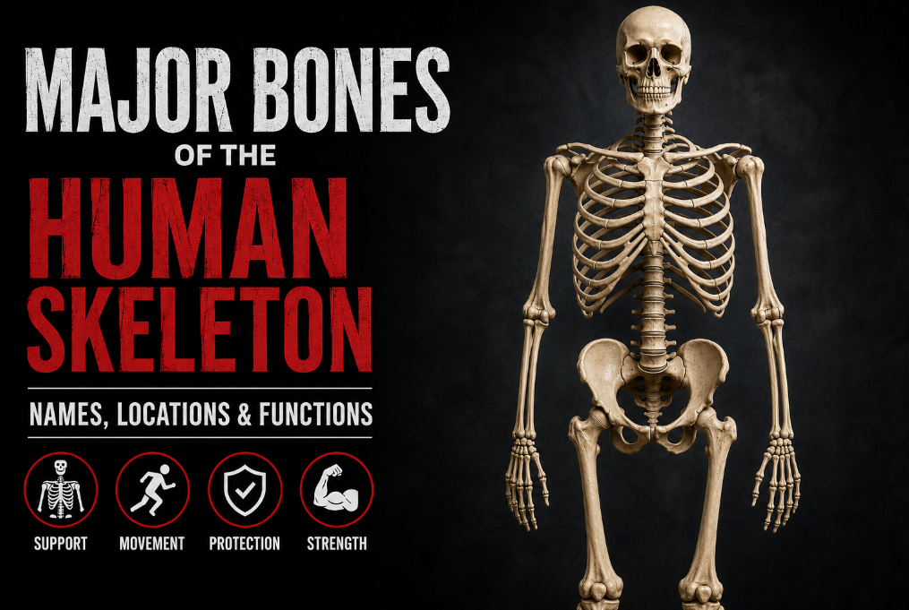
Understanding the major bones of the human skeleton is essential for anyone who wants to learn anatomy, improve exercise technique, or better understand how the body moves. Whether you’re performing squats, bench presses, or deadlifts, knowing where key bones such as the clavicle, scapula, pelvis, and femur are located can help you follow coaching cues more confidently and appreciate the mechanics behind every movement.
Although the adult human skeleton contains 206 bones, only a relatively small number play a dominant role in posture, movement, joint stability, and athletic performance. These major bones provide the framework that supports your body, protects vital organs, and serves as attachment points for muscles, allowing you to generate strength and move efficiently.
In this guide, you’ll learn the names, locations, and primary functions of the major bones of the human skeleton, along with why each one matters for lifters, athletes, healthcare students, and anyone interested in human anatomy. For a deeper look at how these bones function together as a complete system, see our companion guide to the human skeletal system.
Quick Facts
Human Skeleton Overview
The major bones of the human skeleton form a strong but adaptable framework that supports the body, protects vital organs, and allows movement. Although the adult skeleton typically contains 206 bones, these bones do not function independently. They are connected at joints and work together with muscles, tendons, ligaments, cartilage, and other connective tissues to produce controlled movement.
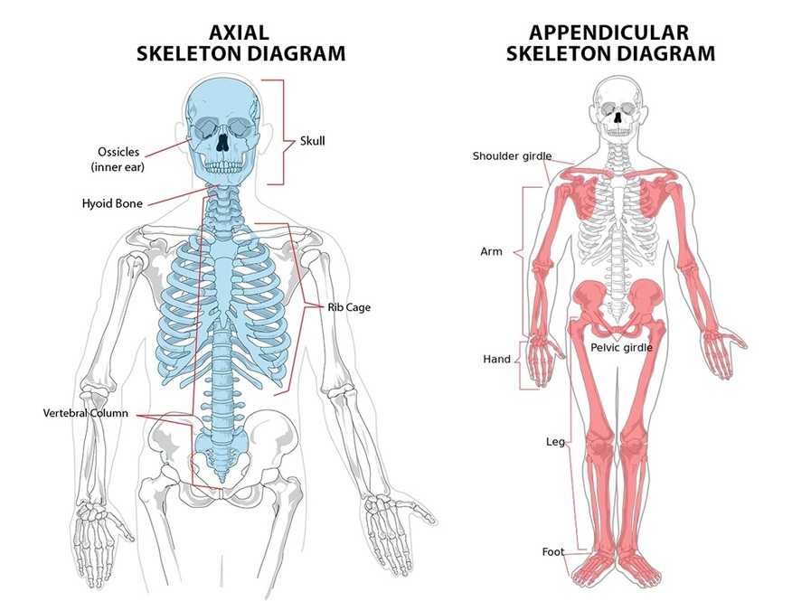
For anatomical study, the skeleton is divided into two main sections: the axial skeleton and the appendicular skeleton. The axial skeleton forms the body’s central axis and includes the skull, vertebral column, ribs, and sternum. Its primary roles are to support, protect, and maintain the posture of the brain, spinal cord, heart, and lungs.
The appendicular skeleton includes the shoulder girdles, upper limbs, pelvic girdle, and lower limbs. These bones are primarily responsible for movement and for transferring force between the upper and lower body. During exercises such as squats, deadlifts, presses, rows, and pull-ups, the bones of the appendicular skeleton move at their joints, while the axial skeleton provides stability and structural support.
The skeleton is therefore more than a collection of rigid bones. It is a coordinated mechanical system that provides leverage, absorbs and transfers force, and creates the structural foundation for nearly every movement you perform.
Axial vs. Appendicular Skeleton
Before exploring the major bones of the human skeleton, it’s important to understand how the skeleton is organized. Anatomists divide the human skeleton into two major sections: the axial skeleton and the appendicular skeleton. Each serves a distinct purpose: one primarily provides stability and protection, and the other enables movement.
The axial skeleton forms the body’s central axis and consists of 80 bones, including the skull, vertebral column, ribs, and sternum. Its primary functions are to support the head and trunk, maintain posture, and protect the brain, spinal cord, heart, and lungs.
The appendicular skeleton consists of 126 bones and includes the shoulder girdles, upper limbs, pelvic girdle, and lower limbs. These bones create the body’s lever system, allowing muscles to generate movement, transfer force, and perform everyday activities as well as athletic movements such as squats, presses, pulls, jumps, and running.
Together, the axial and appendicular skeletons form a complete structural framework that balances stability with mobility, enabling efficient human movement.
| Feature | Axial Skeleton | Appendicular Skeleton |
|---|---|---|
| Number of Bones | 80 | 126 |
| Primary Role | Support and protection | Movement and force production |
| Includes | Skull, vertebral column, ribs, sternum | Shoulder girdles, upper limbs, pelvic girdle, lower limbs |
| Main Function | Protects vital organs and maintains posture | Enables movement and locomotion |
| Importance in Training | Provides stability during lifts | Produces movement and transfers force |
Major Bones of the Human Skeleton by Region
The major bones of the human skeleton can be grouped into different regions of the body based on their location and function. While the skeleton contains 206 bones in total, only a relatively small number are considered major bones because they play key roles in supporting body weight, protecting vital organs, forming major joints, and enabling movement.
The table below provides a quick overview of the most important bones, their locations, and their primary functions. More detailed descriptions of each bone are provided in the following sections.
| Body Region | Major Bone(s) | Primary Function |
|---|---|---|
| Head | Skull | Protects the brain and supports the face |
| Shoulder | Clavicle, Scapula | Connects the upper limb to the trunk and stabilizes the shoulder |
| Chest | Sternum, Ribs | Protect the heart and lungs |
| Spine | Vertebral Column | Supports the body and protects the spinal cord |
| Upper Arm | Humerus | Forms the shoulder and elbow joints |
| Forearm | Radius, Ulna | Allow forearm and wrist movement |
| Hand | Carpals, Metacarpals, Phalanges | Provide hand mobility and grip |
| Pelvis | Pelvic Girdle | Transfers body weight and supports pelvic organs |
| Thigh | Femur | Supports body weight and enables powerful leg movement |
| Knee | Patella | Protects the knee joint and improves quadriceps leverage |
| Lower Leg | Tibia, Fibula | Provide stability and support during standing and walking |
| Foot | Tarsals, Metatarsals, Phalanges | Support balance, locomotion, and force transfer |
Skull

The skull is the body’s most superior bony structure and one of the major bones of the human skeleton. It consists of 22 bones that together form the framework of the head. These bones are divided into the cranial bones, which enclose and protect the brain, and the facial bones, which shape the face and support the eyes, nose, and mouth. Except for the mandible (lower jaw), most skull bones are joined by immovable fibrous joints called sutures.
Location
The skull sits at the top of the vertebral column and forms the head. It articulates with the first cervical vertebra (atlas), allowing movements such as nodding and head rotation.
Primary Functions
- Protects the brain from external injury.
- Supports the structures of the face.
- Houses the organs of vision, hearing, smell, and taste.
- Provides attachment sites for muscles involved in chewing, facial expression, and head movement.
Why It Matters for Lifters
Although the skull itself doesn’t generate movement, its position directly affects head posture, cervical spine alignment, and lifting mechanics. Maintaining a neutral head position during exercises such as squats, deadlifts, and overhead presses helps preserve proper spinal alignment and reduces unnecessary stress on the neck.
Clavicle (Collarbone)
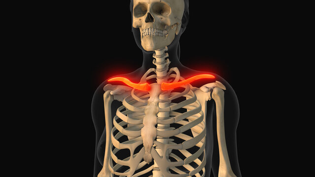
The clavicle, commonly known as the collarbone, is a long, S-shaped bone that connects the upper limb to the trunk. Together with the scapula, it forms the shoulder girdle and plays a crucial role in shoulder mobility and stability. The clavicle is also unique as the only long bone in the human body that lies horizontally.
Location
The clavicle extends from the upper part of the sternum (manubrium) to the acromion of the scapula. It is located just beneath the skin at the front of the shoulder, making it one of the easiest bones to see and feel on the body.
Primary Functions
- Connects the upper limb to the axial skeleton.
- Helps stabilize the shoulder during arm movements.
- Acts as a strut that positions the shoulder away from the chest, allowing a greater range of motion.
- Transmits forces from the upper limb to the trunk.
- Protects important nerves and blood vessels that pass beneath it.
Why It Matters for Lifters
The clavicle plays an important role in nearly every upper-body exercise, including the bench press, overhead press, pull-up, and push-up. Proper clavicular movement helps maintain healthy shoulder mechanics, while fractures or limited mobility can significantly reduce upper-body function and training performance.
Scapula (Shoulder Blade)

The scapula, commonly known as the shoulder blade, is a large, flat, triangular bone located on the upper back. As one of the major bones of the human skeleton, it serves as the foundation for shoulder movement by connecting the upper arm to the clavicle and providing attachment sites for numerous muscles. The scapula plays a central role in upper-body strength, stability, and mobility.
Location
Each scapula lies on the posterior side of the rib cage between the second and seventh ribs. It articulates with the clavicle at the acromioclavicular (AC) joint and with the humerus at the glenohumeral (shoulder) joint, forming part of the highly mobile shoulder complex.
Primary Functions
- Connects the upper arm to the shoulder girdle.
- Provides attachment sites for more than 15 muscles, including the rotator cuff, trapezius, rhomboids, and serratus anterior.
- Stabilizes the shoulder during upper-body movement.
- Allows smooth arm elevation, rotation, and overhead motion.
- Helps transmit forces between the upper limb and the trunk.
Why It Matters for Lifters
The scapula is one of the most important bones for resistance training. Proper scapular movement and stability improve performance during the bench press, overhead press, pull-ups, rows, and push-ups, while also helping reduce the risk of shoulder injuries. Learning how to retract, depress, upwardly rotate, and stabilize the scapula is fundamental for efficient lifting mechanics and long-term shoulder health.
Sternum (Breastbone)
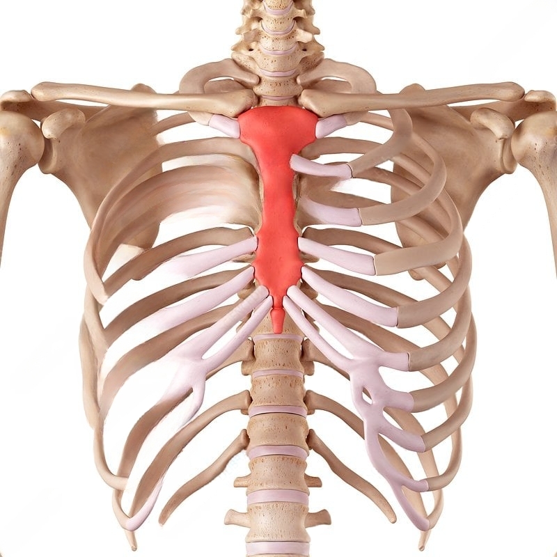
The sternum, commonly known as the breastbone, is a long, flat bone located in the center of the chest. As one of the major bones of the human skeleton, it forms the front of the rib cage and serves as an important attachment point for the ribs, clavicles, and several chest and neck muscles. Its central position makes it essential for protecting vital organs and maintaining upper-body stability.
Location
The sternum lies along the midline of the anterior chest wall. It consists of three parts — the manubrium, body, and xiphoid process — and articulates with both clavicles and the first seven pairs of ribs through costal cartilages.
Primary Functions
- Protects the heart, lungs, and major blood vessels.
- Forms the anterior portion of the rib cage.
- Connects the clavicles to the axial skeleton.
- Provides attachment sites for the ribs and several muscles of the chest, shoulders, neck, and abdomen.
- Helps maintain the stability and structure of the thorax.
Why It Matters for Lifters
The sternum serves as the central anchor for the chest and shoulder girdle during upper-body exercises. Movements such as the bench press, push-up, dips, and cable fly rely on muscles that originate from or attach near the sternum. Understanding its position also helps lifters use coaching cues like “lift your sternum” or “keep your chest up,” which promote better posture and spinal alignment during many compound lifts.
Ribs (Rib Cage)
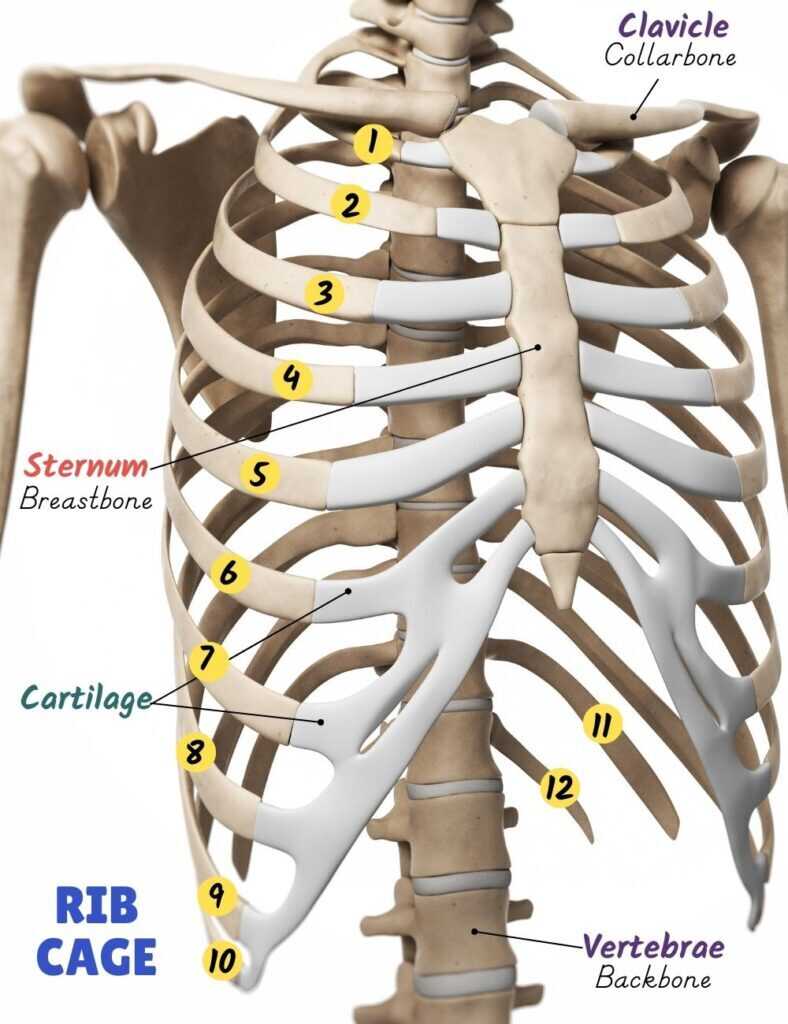
The ribs are long, curved bones that form the rib cage, a protective framework surrounding the heart, lungs, and other vital structures within the thoracic cavity. Together with the sternum and thoracic vertebrae, the ribs create a strong yet flexible cage that protects internal organs while allowing the chest to expand and contract during breathing.
Location
The adult human rib cage consists of 12 pairs (24 ribs) that extend from the thoracic vertebrae toward the front of the chest. The first seven pairs are known as true ribs because they attach directly to the sternum through their own costal cartilages. The next three pairs (false ribs) attach indirectly via the cartilage of the rib above, while the final two pairs (floating ribs) have no anterior attachment to the sternum.
Primary Functions
- Protect the heart, lungs, and major blood vessels.
- Form the framework of the thoracic cavity.
- Support breathing by allowing the chest to expand and contract.
- Provide attachment sites for numerous muscles involved in respiration, posture, and trunk movement.
- Help absorb and distribute forces applied to the upper body.
Why It Matters for Lifters
A stable and properly positioned rib cage contributes to efficient breathing, core stability, and spinal alignment during resistance training. Maintaining an appropriate rib cage position is particularly important during exercises such as the squat, deadlift, overhead press, and bench press, where effective bracing helps transfer force and protect the spine. Understanding rib cage mechanics can also help lifters avoid excessive rib flare, which may compromise posture and lifting technique.
Vertebral Column (Spine)
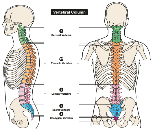
The vertebral column, also known as the spine or backbone, is the central supporting structure of the human body. The human skeleton consists of 33 vertebrae, connected by intervertebral discs and ligaments to provide both stability and flexibility. The spine supports the head and trunk while protecting the delicate spinal cord, making it essential for posture, movement, and overall function.
Location
The vertebral column extends from the base of the skull to the pelvis along the body’s midline. It is divided into five regions: 7 cervical vertebrae, 12 thoracic vertebrae, 5 lumbar vertebrae, 5 fused sacral vertebrae (forming the sacrum), and 4 fused coccygeal vertebrae (forming the coccyx).
Primary Functions
- Supports the weight of the head, neck, and trunk.
- Protects the spinal cord and spinal nerves.
- Maintains upright posture and body alignment.
- Allows movements such as bending, twisting, and extension.
- Transfers forces between the upper and lower body.
Why It Matters for Lifters
The spine is one of the most important structures in resistance training because nearly every compound exercise depends on proper spinal alignment. Maintaining a neutral spine during exercises such as the squat, deadlift, overhead press, bent-over row, and Romanian deadlift helps distribute loads efficiently, improve force transfer, and reduce the risk of spinal injuries. A strong and stable spine also provides the foundation for safe and effective movement under heavy loads.
Humerus (Upper Arm Bone)

The humerus is the long bone of the upper arm that extends from the shoulder to the elbow. As one of the major bones of the human skeleton, it forms the connection between the shoulder girdle and the forearm, making it essential for nearly every upper-body movement. The humerus also serves as an attachment site for many muscles responsible for lifting, pushing, pulling, and throwing.
Location
The humerus is located between the shoulder and elbow. Its rounded head articulates with the scapula to form the glenohumeral (shoulder) joint, while its distal end articulates with the radius and ulna to form the elbow joint.
Primary Functions
- Connects the shoulder to the forearm.
- Forms the shoulder and elbow joints.
- Acts as a lever for upper-limb movement.
- Provides attachment sites for numerous muscles, including the deltoid, pectoralis major, latissimus dorsi, biceps brachii, triceps brachii, and brachialis.
- Transmits forces between the upper arm and forearm.
Why It Matters for Lifters
The humerus is involved in virtually every upper-body exercise, including the bench press, overhead press, pull-up, lat pulldown, row, dip, and biceps curl. Its position influences shoulder mechanics, range of motion, and lifting efficiency. Understanding how the humerus moves within the shoulder joint can also help lifters improve technique and reduce the risk of shoulder injuries.
Radius and Ulna (Forearm Bones)
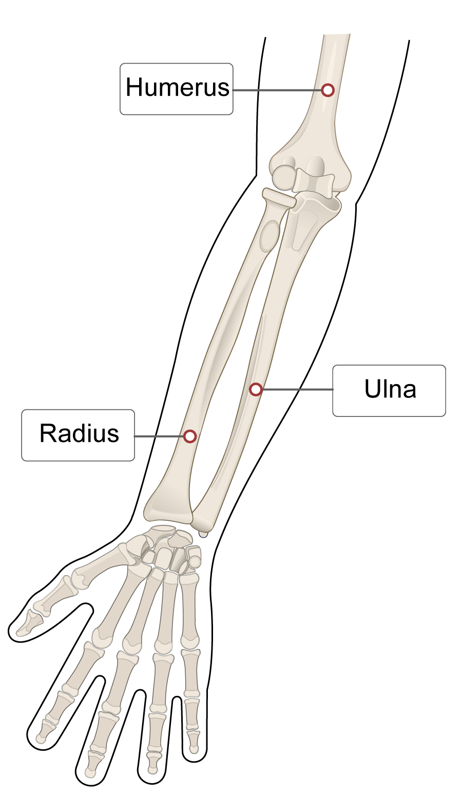
The radius and ulna are the two long bones of the forearm that work together to provide strength, stability, and a wide range of arm and hand movements. As major bones of the human skeleton, they form the connection between the elbow and wrist, allowing the forearm to rotate while supporting gripping, lifting, and carrying activities.
Location
The radius and ulna extend from the elbow to the wrist. In the anatomical position (palms facing forward), the radius is located on the thumb side of the forearm, while the ulna lies on the little finger side. These bones articulate with each other at both ends, allowing the forearm to rotate during pronation and supination.
Primary Functions
- Form the forearm and connect the elbow to the wrist.
- Allow forearm rotation during pronation and supination.
- Stabilize the elbow and wrist joints.
- Transfer forces between the hand and upper arm.
- Provide attachment sites for muscles that move the wrist, hand, and fingers.
Why It Matters for Lifters
The radius and ulna are heavily involved in nearly every upper-body exercise that requires gripping a barbell, dumbbell, cable handle, or pull-up bar. They play an essential role in exercises such as biceps curls, rows, pull-ups, deadlifts, bench presses, and farmer’s carries. Understanding forearm rotation also helps lifters choose the most appropriate grip — such as pronated, supinated, or neutral — for different exercises and training goals.
Hand Bones (Carpals, Metacarpals, and Phalanges)
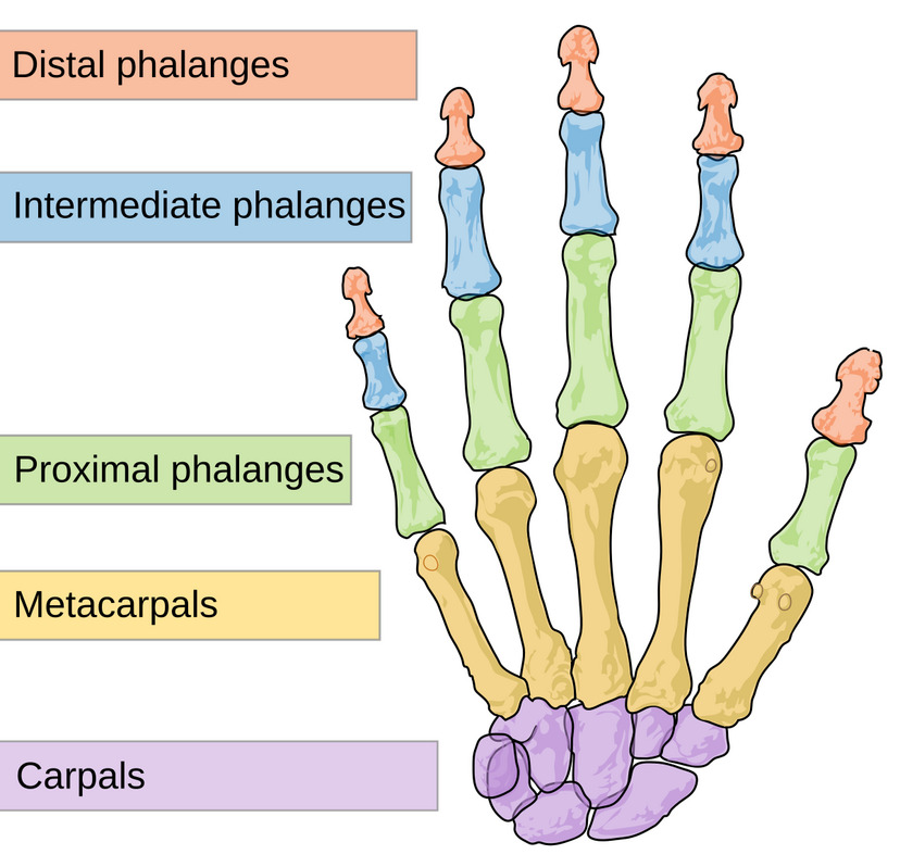
The bones of the hand consist of 27 bones that work together to provide exceptional mobility, dexterity, and grip strength. As major bones of the human skeleton, they enable everything from fine motor tasks, such as writing and typing, to powerful gripping movements required for resistance training and sports. The hand is divided into three groups of bones: the carpals, metacarpals, and phalanges.
Location
The hand contains 8 carpal bones that form the wrist, 5 metacarpal bones that make up the palm, and 14 phalanges that form the fingers and thumb. Together, these bones create a highly mobile structure capable of both precision and forceful gripping.
Primary Functions
- Form the framework of the wrist, palm, and fingers.
- Enable gripping, pinching, and fine motor control.
- Support a wide range of hand and finger movements.
- Transfer forces between the hand and forearm.
- Provide attachment sites for muscles, tendons, and ligaments that control hand function.
Why It Matters for Lifters
Strong and healthy hand bones are essential for maintaining a secure grip during exercises such as deadlifts, pull-ups, rows, farmer’s carries, and shrugs. Grip strength often becomes the limiting factor in heavy pulling exercises, making the hand an important link in the kinetic chain. Maintaining wrist stability and hand strength can also improve lifting performance while reducing the risk of overuse injuries.
Pelvic Girdle (Pelvis)

The pelvic girdle, commonly referred to as the pelvis, is a strong, basin-shaped bony structure that connects the spine to the lower limbs. As one of the major bones of the human skeleton, it provides a stable foundation for standing, walking, and nearly every lower-body movement. The pelvis also protects several vital organs and plays a key role in transferring forces between the upper and lower body.
Location
The pelvic girdle is located at the base of the trunk between the lumbar spine and the femurs. It consists of two hip bones (ossa coxae), each formed by the fusion of the ilium, ischium, and pubis. Together with the sacrum and coccyx, these bones form the bony pelvis.
Primary Functions
- Connects the axial skeleton to the lower limbs.
- Transfers body weight from the spine to the legs.
- Supports and protects the pelvic organs, including the bladder and reproductive organs.
- Provides attachment sites for numerous muscles of the hips, thighs, abdomen, and pelvic floor.
- Contributes to balance, posture, and efficient force transfer during movement.
Why It Matters for Lifters
The pelvis is one of the most important structures in strength training because it serves as the foundation for lower-body power production. Exercises such as the squat, deadlift, hip thrust, lunge, and Olympic lifts all rely on proper pelvic alignment for efficient force transfer and spinal stability. Learning to maintain a neutral pelvis can improve lifting mechanics, enhance performance, and reduce stress on the lower back and hips.
Femur (Thigh Bone)
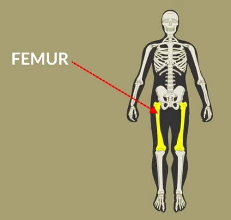
The femur, commonly known as the thigh bone, is the longest, largest, and strongest bone in the human body. As one of the major bones of the human skeleton, it forms the upper part of the lower limb and plays a central role in weight-bearing, locomotion, and force production. The femur also serves as the primary connection between the pelvis and the lower leg, making it indispensable for both everyday activities and athletic performance.
Location
The femur extends from the hip to the knee. Its rounded head fits into the acetabulum of the pelvis to form the hip joint, while its distal end articulates with the tibia and patella to form the knee joint.
Primary Functions
- Supports the body’s weight during standing and movement.
- Connects the pelvis to the lower leg.
- Forms the hip and knee joints.
- Acts as a powerful lever for walking, running, jumping, and lifting.
- Provides attachment sites for many of the body’s largest muscles, including the gluteal muscles, quadriceps, hamstrings, and adductors.
Why It Matters for Lifters
The femur is one of the most important bones in strength training because it plays a major role in virtually every lower-body exercise. Movements such as the squat, deadlift, leg press, lunge, Bulgarian split squat, and hip thrust all depend on efficient force transfer through the femur. Its length and angle also influence squat depth, stance width, and overall lifting mechanics, making femoral anatomy an important factor in exercise technique.
Patella (Kneecap)
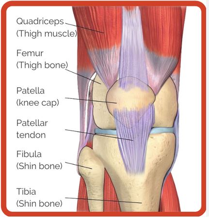
The patella, commonly known as the kneecap, is the largest sesamoid bone in the human body. As one of the major bones of the human skeleton, it is embedded within the quadriceps tendon and protects the front of the knee joint. Beyond its protective role, the patella improves the mechanical efficiency of the quadriceps muscles, making knee extension more powerful.
Location
The patella is located at the front of the knee, where it sits within the quadriceps tendon. It articulates with the femur at the patellofemoral joint and connects to the tibia through the patellar ligament.
Primary Functions
- Protects the front of the knee joint from direct impact.
- Increases the leverage and mechanical advantage of the quadriceps muscles.
- Improves the efficiency of knee extension.
- Helps guide the quadriceps tendon during knee motion.
- Contributes to knee stability during movement.
Why It Matters for Lifters
The patella plays a critical role in nearly every lower-body exercise involving knee extension, including the squat, leg press, lunge, step-up, leg extension, and Bulgarian split squat. Proper patellar tracking is essential for healthy knee mechanics, while poor alignment or excessive loading may contribute to conditions such as patellofemoral pain syndrome. Understanding the patella’s function helps lifters appreciate the importance of proper technique, balanced muscle development, and controlled movement patterns.
Tibia and Fibula (Lower Leg Bones)
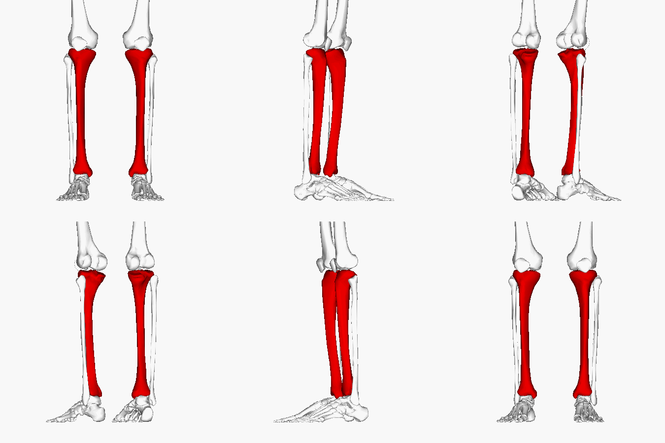
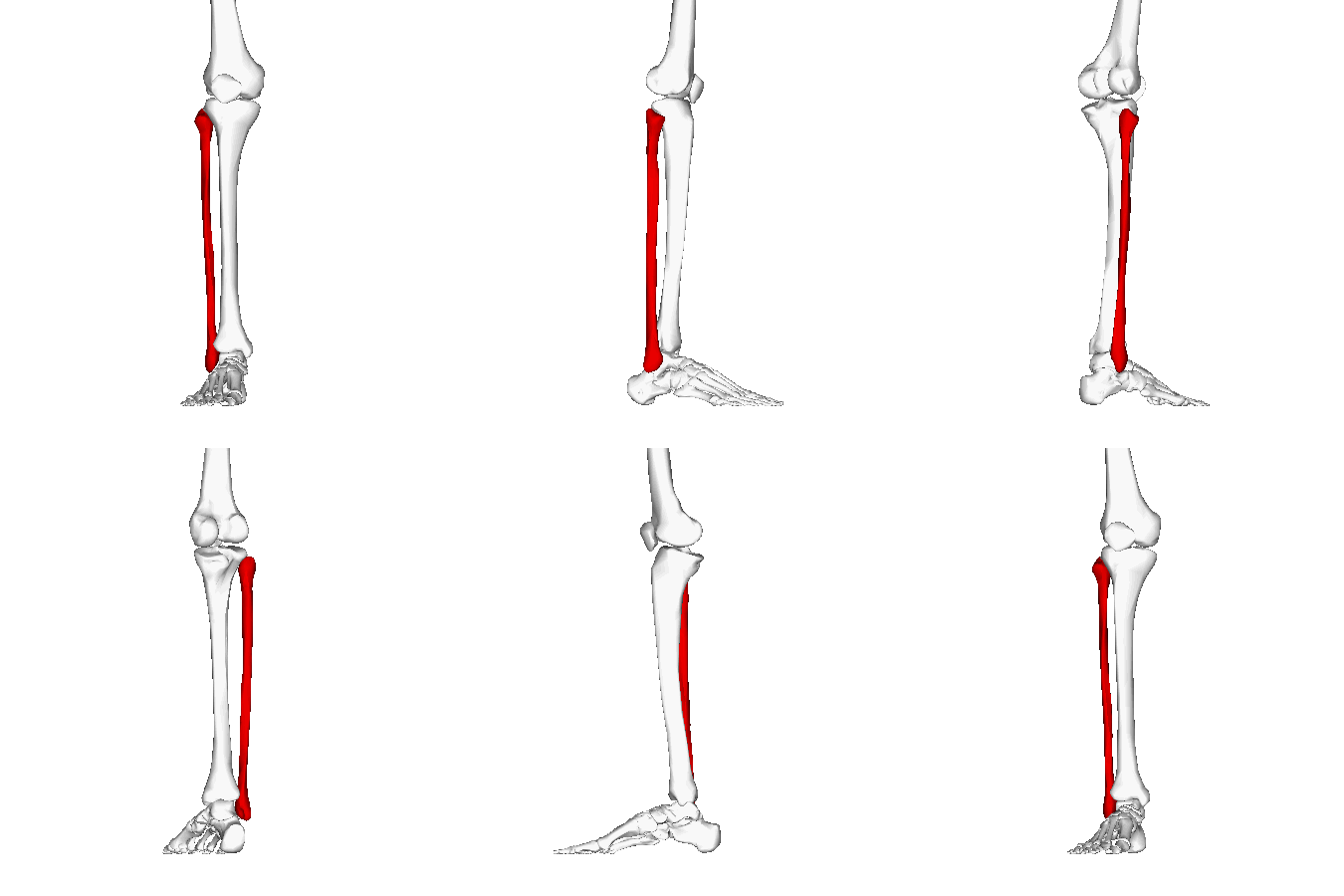
The tibia and fibula are the two long bones of the lower leg that work together to provide support, stability, and movement. As major bones of the human skeleton, they connect the knee to the ankle and play essential roles in weight-bearing, balance, and locomotion. Although they function as a pair, each bone has distinct anatomical and mechanical responsibilities.
Location
The tibia, or shinbone, is the larger and stronger of the two bones and is located on the medial (inner) side of the lower leg. It bears most of the body’s weight and forms both the knee and ankle joints. The fibula is the thinner bone located on the lateral (outer) side of the leg. While it bears relatively little body weight, it provides important attachment sites for muscles and contributes significantly to ankle stability.
Primary Functions
- Support body weight during standing, walking, and running.
- Form the lower portion of the knee joint and the upper portion of the ankle joint.
- Provide stability to the lower leg and ankle.
- Serve as attachment sites for muscles that control movement of the knee, ankle, and foot.
- Transfer forces between the foot and the femur during weight-bearing activities.
Why It Matters for Lifters
The tibia and fibula are essential for virtually every lower-body exercise. Movements such as the squat, deadlift, leg press, calf raise, lunge, and jump all rely on these bones to transmit forces safely between the ground and the rest of the body. Proper lower-leg alignment also influences knee and ankle mechanics, helping improve lifting efficiency while reducing the risk of stress fractures, shin splints, and other overuse injuries.
Foot Bones (Tarsals, Metatarsals, and Phalanges)

The bones of the foot consist of 26 bones that work together to provide stability, balance, shock absorption, and propulsion during movement. As part of the major bones of the human skeleton, these bones form a strong yet flexible structure capable of supporting the body’s weight while adapting to different surfaces and movement demands. The foot is divided into three groups of bones: the tarsals, metatarsals, and phalanges.
Location
The foot contains 7 tarsal bones, which form the ankle and heel, 5 metatarsal bones, which make up the midfoot, and 14 phalanges, which form the toes. Together, these bones form the longitudinal and transverse arches of the foot, which help distribute body weight and absorb impact during standing, walking, running, and jumping.
Primary Functions
- Support the body’s weight during standing and movement.
- Maintain balance and stability.
- Absorb shock during walking, running, and jumping.
- Provide leverage for propulsion during gait.
- Form the structural arches of the foot.
- Provide attachment sites for muscles, tendons, and ligaments that control foot and toe movement.
Why It Matters for Lifters
The bones of the foot provide the foundation for nearly every standing exercise. Movements such as the squat, deadlift, lunge, calf raise, Olympic lifts, and farmer’s carry all depend on a stable foot to transfer force efficiently from the ground through the rest of the body. Good foot stability, arch control, and proper weight distribution can improve lifting performance, balance, and overall movement mechanics while reducing the risk of lower-extremity injuries.
Bones Every Lifter Should Know
While every bone in the body serves an important purpose, some have a much greater impact on exercise performance, lifting technique, and injury prevention than others. Understanding these key bones can help you follow coaching cues more effectively, improve movement mechanics, and reduce unnecessary stress on your joints during resistance training.
| Bone | Why It Matters for Lifters | Common Exercises |
|---|---|---|
| Scapula | Stabilizes the shoulder and allows efficient upper-body movement. | Bench press, pull-ups, rows, overhead press |
| Clavicle | Connects the upper limb to the trunk and supports shoulder mechanics. | Bench press, push-ups, dips |
| Vertebral Column | Maintains spinal stability and transfers force throughout the body. | Squat, deadlift, bent-over row |
| Pelvis | Forms the foundation for lower-body power and hip stability. | Squat, deadlift, hip thrust |
| Femur | Primary weight-bearing bone responsible for powerful hip and knee movement. | Squat, leg press, lunge |
| Patella | Improves quadriceps leverage during knee extension. | Squat, leg extension, step-up |
| Tibia | Bears most of the body’s weight and stabilizes the knee and ankle. | Squat, calf raise, walking lunge |
| Hand Bones | Provide grip strength and control during lifting. | Deadlift, pull-up, farmer’s carry |
Interesting Facts About the Human Skeleton
The human skeleton is far more dynamic than many people realize. Although bones appear rigid and unchanging, they are living tissues that constantly adapt to mechanical stress, repair damage, and support nearly every movement you perform. Here are some fascinating facts about the major bones of the human skeleton and the skeletal system as a whole.
Largest, Longest, and Smallest Bones
- Longest bone: The femur (thigh bone), measuring approximately 48 cm (19 in) in an average adult.
- Largest bone: The femur, which is also the body’s heaviest and strongest bone.
- Smallest bone: The stapes, located in the middle ear, measuring only about 3 mm in length.
Babies Have More Bones Than Adults
Newborn babies have approximately 270 bones. As they grow, many of these bones gradually fuse together, resulting in the 206 bones found in the average adult skeleton.
Bones Are Living Tissue
Bones are continuously remodeled throughout life by specialized cells. This process repairs microscopic damage, helps maintain bone strength, and allows the skeleton to adapt to physical activity and mechanical loading.
Bones Store Essential Minerals
In addition to providing structural support, bones act as the body’s primary storage site for calcium and phosphorus. These minerals can be released into the bloodstream when needed to support normal muscle contraction, nerve function, and other physiological processes.
The Skeleton Produces Blood Cells
Inside many bones is red bone marrow, where millions of new red blood cells, white blood cells, and platelets are produced every day through a process known as hematopoiesis.
Bones Become Stronger with Resistance Training
Weight-bearing exercise and resistance training stimulate bone remodeling, helping maintain or increase bone mineral density over time. This is one of the many reasons strength training is recommended throughout adulthood.
Why Learning the Major Bones of the Human Skeleton Matters
Learning the major bones of the human skeleton is about much more than memorizing anatomical terms. Whether you’re a student, healthcare professional, personal trainer, or fitness enthusiast, understanding where the body’s major bones are located and how they function provides a stronger foundation for learning human movement and improving exercise performance.
Knowing the relationship between bones, joints, and muscles makes it easier to understand exercise technique, follow coaching instructions, and recognize how the body generates and transfers force. It can also help you identify common movement errors, appreciate the importance of proper posture, and better understand the mechanisms behind many musculoskeletal injuries.
For lifters and athletes, skeletal anatomy provides valuable insight into biomechanics. Understanding how the scapula influences shoulder stability, how the pelvis transfers force between the upper and lower body, or how the femur affects squat mechanics allows you to train with greater confidence and efficiency.
As your knowledge of anatomy grows, you’ll begin to see the human skeleton not simply as a collection of bones, but as the structural framework that makes every movement possible — from everyday activities to the heaviest lifts in the gym. Want to see how all these bones connect visually? Try our interactive human skeleton tool to explore each one in detail.
Key Takeaways
- The adult human skeleton contains 206 bones, divided into the axial skeleton (80 bones) and appendicular skeleton (126 bones).
- The major bones of the human skeleton provide structural support, protect vital organs, and serve as attachment points for muscles, enabling efficient movement.
- The femur is the body’s longest and largest bone, while the stapes in the middle ear is the smallest.
- Bones such as the vertebral column, pelvis, scapula, clavicle, humerus, femur, tibia, and fibula play especially important roles in posture, mobility, and resistance training.
- Understanding the location and function of the body’s major bones makes it easier to follow exercise instructions and improve lifting technique.
- Learning skeletal anatomy can help reduce injury risk and deepen your understanding of human movement and biomechanics.
What are the major bones of the human skeleton?
The major bones of the human skeleton include the skull, clavicle, scapula, sternum, ribs, vertebral column, humerus, radius, ulna, hand bones, pelvis, femur, patella, tibia, fibula, and foot bones. Together, they provide structural support, protect vital organs, and enable movement by serving as attachment points for muscles.
How many bones are in the human skeleton?
A healthy adult human skeleton contains 206 bones. Infants are born with approximately 270 bones, many of which gradually fuse together during growth and development.
What is the largest bone in the human body?
The femur, or thigh bone, is the largest, longest, and strongest bone in the human body. It supports much of the body's weight and plays a vital role in walking, running, jumping, and strength-training exercises such as squats and deadlifts.
What is the smallest bone in the human body?
The stapes, located in the middle ear, is the smallest bone in the human body. It measures only about 3 millimeters (0.12 inches) in length and helps transmit sound vibrations to the inner ear.
Which bones bear the most body weight?
The femur, tibia, and the bones of the pelvis bear the greatest amount of body weight during standing, walking, running, and lifting. These bones are specifically adapted to withstand high compressive forces while maintaining stability and efficient movement.
Which bones are most important for strength training?
Although every bone contributes to movement, lifters should be especially familiar with the scapula, clavicle, vertebral column, pelvis, femur, tibia, patella, and hand bones. These structures play key roles in stability, force transfer, posture, and lifting mechanics.
What is the difference between the axial and appendicular skeleton?
The axial skeleton consists of the skull, vertebral column, ribs, and sternum, primarily supporting posture and protecting vital organs. The appendicular skeleton includes the bones of the arms, legs, shoulders, and pelvis, allowing movement and locomotion.
Are bones living tissue?
Yes. Bones are living connective tissue that continuously remodel throughout life. Specialized cells constantly break down old bone while forming new bone, allowing the skeleton to repair microscopic damage and adapt to mechanical stress.
Does strength training make bones stronger?
Yes. Regular resistance training and other weight-bearing exercises stimulate bone remodeling, helping maintain or increase bone mineral density. When combined with adequate nutrition, strength training supports lifelong skeletal health.
Why is learning bone anatomy important?
Understanding bone anatomy helps you better understand human movement, exercise technique, biomechanics, posture, and injury prevention. It also makes it easier to follow coaching cues, study anatomy, and appreciate how muscles and joints work together to produce movement.
