

Every squat, deadlift, bench press, or pull-up looks like a simple move from point A to point B. It isn’t. Behind that single rep sits a coordinated system: your nervous system fires a signal, a muscle develops tension, a tendon transmits that tension to a bone, and a joint converts it into motion. Miss any link in that chain and the rep either doesn’t happen or happens badly.
That entire force-producing system — from the smallest contractile protein inside a muscle fiber to the largest muscle in your body — is what we call the muscular system. It is not a pile of “meat” that gets bigger when you train. It’s a living organ system that generates force, controls posture, stabilizes joints, and physically changes its own structure in response to how you load it.
This guide is built specifically for lifters, coaches, and athletes rather than medical students. We’ll cover what the muscular system is, how it’s organized from whole muscle down to actin and myosin, how it actually contracts, how it works together with your nervous and skeletal systems, and — most importantly — what happens to it during and after a strength-training session. Along the way, we’ll point you toward our more detailed guides on specific muscles, fiber types, and growth mechanisms, so this article stays a map rather than an encyclopedia.
QUICK ANSWER: The muscular system is the organ system responsible for producing force and movement in the human body. It includes three tissue types — skeletal, cardiac, and smooth muscle — but for lifters, skeletal muscle is the primary focus, since it’s the voluntary tissue that attaches to bone and drives every rep you perform. Skeletal muscle works together with the nervous system (which activates it) and the skeletal system (which it moves), and it’s also one of the most adaptable tissues in the body: resistance training reshapes its size, fiber composition, and metabolic capacity over time.
What Is the Muscular System?
The muscular system is one of the eleven organ systems of the human body. Its job is to generate the mechanical force needed for movement, to stabilize joints and posture, and to support several less obvious functions we’ll get into later — heat production, circulation, and even metabolism.
The Muscular System Is More Than Your Skeletal Muscles
Most lifters use “muscular system” and “skeletal muscle” as interchangeable terms, but that’s only partly accurate. The muscular system technically includes all three types of muscle tissue in the body — skeletal, cardiac, and smooth — even though only one of those (skeletal muscle) is what you’re training when you lift weights.
How Many Muscles Are in the Human Body?
No single, universally agreed-upon number exists. Anatomy sources commonly cite a figure in the region of 600, but the true count depends heavily on how individual muscles, heads, and slips are classified — some muscles are counted as one, others split into separate heads, and anatomical variation between individuals adds further inconsistency. What matters more for training purposes isn’t landing on an exact count — it’s understanding that these muscles are organized into functional groups that cooperate rather than working as hundreds of independent units. Each of these muscles also has a name that isn’t random — see our guide on how skeletal muscles are named if you’ve ever wondered why muscle names look the way they do.”
Muscles as Organs, Not Just Contractile Tissue
A skeletal muscle isn’t simply a bundle of contractile fibers. It’s an organ in its own right, made up of muscle fibers, connective tissue, blood vessels, and nerves, all working together. This matters practically: when a muscle is injured, it’s rarely just the contractile fibers that are affected — the connective tissue and vascular supply matter just as much for recovery and force transmission.
| Feature | Basic Fact |
| Main tissue types | Skeletal, cardiac, smooth |
| Voluntary muscle | Primarily skeletal muscle |
| Primary movement role | Force production and transmission |
| Connection to skeleton | Primarily through tendons |
| Controlled by | The nervous system |
| Major training adaptation | Hypertrophy plus neural and metabolic adaptations |
What Are the Three Types of Muscle Tissue?
Skeletal Muscle
Skeletal muscle is striated (it has a banded appearance under a microscope), primarily voluntary, and attaches to the skeleton via tendons. It’s responsible for movement, posture, joint stability, and force production — which is why it’s the tissue this entire article, and this entire website, is really about.
Cardiac Muscle
Cardiac muscle (myocardium) makes up the walls of the heart. It’s striated like skeletal muscle but involuntary — you don’t consciously decide to contract it — and it’s built for continuous, rhythmic contraction rather than the intermittent, on-demand contractions of skeletal muscle. Endurance training does improve cardiac muscle function (stroke volume, contractile efficiency), which is part of why cardiovascular conditioning matters even for physique-focused lifters.
Smooth Muscle
Smooth muscle lines internal structures like blood vessels, the gastrointestinal tract, and the airways. It’s involuntary and non-striated, and while it’s not something you train directly in the gym, it does respond indirectly to training — for example, vascular smooth muscle adapts to support the increased blood flow demands of trained muscle.
| Feature | Skeletal | Cardiac | Smooth |
| Appearance | Striated | Striated | Non-striated |
| Control | Voluntary | Involuntary | Involuntary |
| Location | Attached to bone | Heart wall | Vessels, organs, airways |
| Trainability | Highly trainable (hypertrophy) | Trainable via cardio | Adapts indirectly |
Why Skeletal Muscle Matters Most for Lifters
From this point on, this guide focuses almost entirely on skeletal muscle, because it’s the only one of the three tissue types you can deliberately reshape through training. Skeletal muscle drives movement, force output, strength, and hypertrophy; it stabilizes your joints under load; it produces the posture you hold between sets; and, less obviously, it plays a real role in whole-body glucose disposal and metabolic health.
Bodybuilder’s Take: For bodybuilding purposes, skeletal muscle is both the machinery that performs the training and the tissue you’re actually trying to change with that training. Every set you perform is simultaneously an act of using the muscular system and an attempt to reshape it — which is exactly why understanding how it’s built and how it adapts isn’t academic trivia, it’s the basis for every programming decision you’ll ever make.
How Is a Skeletal Muscle Organized?
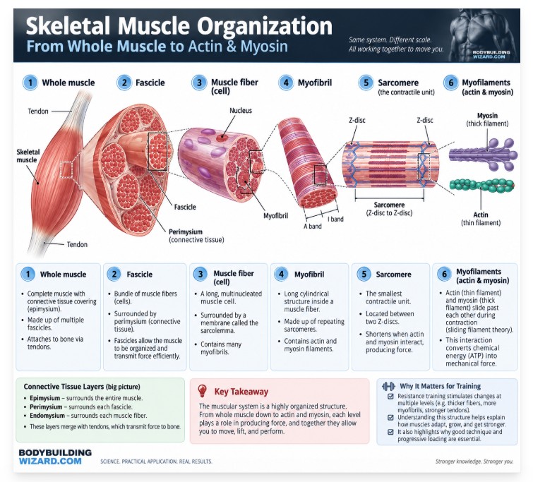
Skeletal muscle is organized hierarchically, from the macroscopic down to the molecular: whole muscle → fascicle → muscle fiber → myofibril → sarcomere → myofilaments (actin and myosin). Understanding this hierarchy is what lets you make sense of concepts like hypertrophy, fiber recruitment, and muscle architecture later in this guide.
Whole Muscle
The whole muscle — your biceps brachii, your gluteus maximus, your pectoralis major — is what you can see and feel. It’s wrapped in a connective-tissue sheath and attaches to bone at each end.
Muscle Fascicles
Inside the whole muscle, fibers are bundled into fascicles — think of them as cables within the larger cable. You can sometimes see the “grain” of a fascicle bundle in raw meat; the same structure exists in human muscle.
Muscle Fibers
Each fascicle contains individual muscle fibers, single elongated muscle cells that can run the entire length of some muscles.
Myofibrils
Inside each muscle fiber are myofibrils — rod-like structures packed with the contractile machinery that actually generates force.
Sarcomeres
Myofibrils are made of repeating units called sarcomeres, which are the basic functional unit of muscle contraction.
Actin and Myosin
Each sarcomere contains two proteins that make contraction possible: actin (thin filaments) and myosin (thick filaments). Their interaction — the sliding filament mechanism — is what we’ll cover in the contraction section below.
We go into fiber types in far more depth in our dedicated guide: Muscle Fibers Explained: Types, Structure, Function & Training.
The Connective Tissue Layers of Skeletal Muscle
This is a part most fitness content skips entirely — but it’s exactly the kind of anatomical depth that separates a resource written by a kinesiologist from a generic blog post. Every level of the muscle hierarchy above is wrapped in its own layer of connective tissue.
Epimysium
The epimysium is the outermost layer and surrounds the entire muscle.
Perimysium
The perimysium wraps each fascicle (bundle of fibers).
Endomysium
The endomysium surrounds each individual muscle fiber.
These three layers aren’t just packaging — they merge at the ends of the muscle to form the tendon, creating a continuous mechanical pathway that transmits force from the contracting fiber all the way to the bone. If any part of this muscle–connective tissue–tendon complex is weak, damaged, or poorly conditioned, force transmission suffers, regardless of how strong the contractile tissue itself is. For a deeper look at how this tissue adapts to training, see our guides on Connective Tissue: Types, Anatomy, Function & Training Adaptation and Tendons and Ligaments: Anatomy, Function & Training Adaptation.
How Muscles Attach to Bones
Tendons and Aponeuroses
Most muscles attach to bone via tendons — dense, cord-like connective tissue. Some broad, flat muscles instead attach via a sheet-like structure called an aponeurosis.
Muscle Origin and Insertion
Every muscle has an origin (the more fixed attachment point) and an insertion (the point that moves when the muscle contracts). Take the biceps brachii: its origin is on the scapula, and its insertion is on the radius in the forearm — which is exactly why contracting the biceps flexes the elbow. The name itself is a clue: ‘bi-‘ signals two heads, and ‘brachii’ places it in the arm — see our full guide to how muscle names are built.
Why Attachment Points Affect Movement
Where a muscle attaches relative to a joint determines what movement it can produce and how much leverage it has. We cover this in far more mechanical detail — including how it affects specific muscles like the biceps and glutes — in Muscle Anatomy Basics: Points of Origin & Insertion, Innervation.
How Does a Muscle Actually Contract?
From Brain Signal to Muscle Contraction
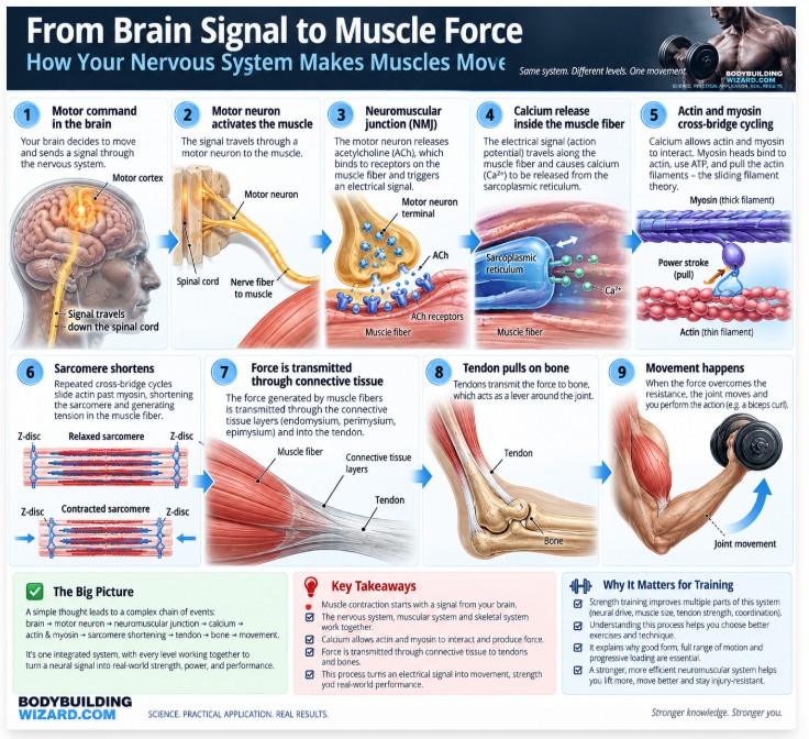
Contraction begins in the brain or spinal cord, travels down a motor neuron, crosses the neuromuscular junction, and triggers muscle fiber activation.
Calcium and Muscle Activation
When the signal arrives, calcium is released inside the muscle fiber, exposing binding sites on the actin filament.
Sliding Filament Theory
Once those binding sites are exposed, myosin heads attach to actin and pull it inward, shortening the sarcomere. This is the sliding filament mechanism — the actin and myosin filaments themselves don’t shorten; they slide past one another.
How Actin and Myosin Produce Force
Each myosin head performs a small “power stroke,” and thousands of these strokes happening simultaneously across a muscle fiber add up to the force you feel when you push a bar off your chest.
ATP and the Cross-Bridge Cycle
Each power stroke consumes ATP. This repeating attach-pull-release-reset sequence is called the cross-bridge cycle, and it’s why muscle contraction is fundamentally an energy-consuming process — one reason fatigue sets in as ATP and other metabolic resources are used up during a set.
How the Muscular, Nervous and Skeletal Systems Work Together
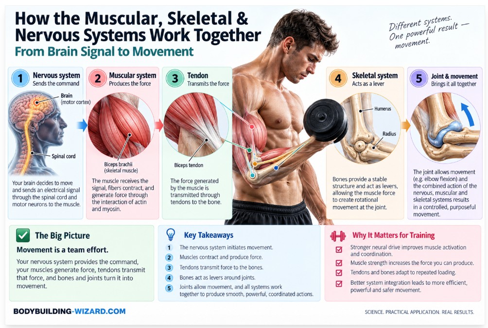
Muscle contraction never happens in isolation. In practice, the nervous system activates the muscle, the muscular system produces force, tendons transmit that force, bones act as levers, and joints permit the resulting motion. Take any one of these away and the movement doesn’t happen — or happens inefficiently and unsafely.
What Happens During a Biceps Curl?
It’s easier to see this system in action with one concrete example. When you curl a dumbbell, a motor command leaves the brain and travels to the motor neurons controlling your elbow flexors; those muscles develop tension; the tension is transmitted through the biceps tendon to the radius; and the resulting torque at the elbow joint overcomes the weight of the dumbbell. Every single rep you’ve ever performed followed this exact sequence.
What Are the Main Functions of the Muscular System?
It’s easy to reduce the muscular system’s job to “movement,” but that undersells what it actually does.
Producing Movement
The most obvious function: skeletal muscle contraction moves the skeleton.
Maintaining Posture and Body Position
Many muscles are working isometrically at any given moment just to keep you upright — this is why postural muscles (like the deep spinal stabilizers) are built for endurance rather than raw power.
Stabilizing Joints
Muscles crossing a joint help hold it in a stable, controlled position, which is part of why weak stabilizer muscles (like the rotator cuff or gluteus medius) show up as instability elsewhere in the kinetic chain.
Producing Heat
Skeletal muscle is an important source of body heat. During exercise, much of the energy released by contracting muscle appears as heat rather than mechanical work, and shivering — rapid, involuntary skeletal-muscle contractions — is one of the body’s main defenses for increasing heat production when it’s cold.
Supporting Circulation and Venous Return
Skeletal muscle contractions around veins, especially in the legs, help push blood back toward the heart against gravity — often called the “skeletal muscle pump.”
Protecting and Supporting Internal Structures
Abdominal wall and pelvic floor muscles help protect and support internal organs.
Metabolic and Endocrine Roles of Skeletal Muscle
This is the part most fitness content leaves out entirely. Skeletal muscle isn’t just tissue that moves weight — it’s one of the body’s largest sites of glucose disposal, meaning more muscle mass generally supports better blood sugar regulation. It also functions as an endocrine, secretory organ: contracting muscle releases signaling molecules called myokines that communicate with other tissues, including fat and the brain. This is part of why resistance training carries benefits that go well beyond aesthetics or strength numbers.
How Do Muscles Produce Different Types of Movement?
Muscles produce force through three basic types of contraction:
| Muscle Action | What Happens | Gym Example |
| Concentric | Muscle produces force while shortening | Rising phase of a biceps curl |
| Eccentric | Muscle produces force while lengthening | Lowering the dumbbell under control |
| Isometric | Tension without appreciable joint movement | Holding a plank or a mid-rep pause |
We break these down in much more detail — including why eccentric-focused training tends to cause more soreness — in Types of Muscle Actions: Isotonic vs. Isometric.
How Muscles Work Together to Produce Movement
A single muscle rarely acts alone. Most joint movements involve several muscles playing different roles at the same time.
Agonists
The agonist (or “prime mover”) is the muscle primarily responsible for producing a given movement. During elbow flexion — a biceps curl, for example — the biceps brachii is the agonist.
Antagonists
The antagonist is the muscle on the opposite side of the joint that lengthens (or controls the movement eccentrically) to allow the agonist to act. In that same elbow-flexion example, the triceps brachii is the antagonist.
Synergists
Synergists assist the agonist, often by fine-tuning the direction of movement or sharing some of the mechanical load. In the bench press, the pectoralis major is the primary mover, while the anterior deltoid and triceps act as synergists that share the load and help control the bar path.
Stabilizers
Stabilizers don’t produce the movement at all — they hold a joint or body segment steady so the prime movers and synergists can work efficiently. In that same bench press, the rotator cuff and scapular musculature act as stabilizers, controlling and bracing the shoulder blade and shoulder joint under load.
This is one of the biggest conceptual gaps in most gym-goers’ understanding of anatomy: no major lift is ever really a “one-muscle exercise.” We’ll be publishing a full breakdown of this concept soon in a dedicated guide on agonist, antagonist, synergist, and stabilizer muscles — for now, keep this framework in mind every time you look at an exercise’s “muscles worked” list.
We break down exactly how that force travels from muscle fiber through connective tissue and tendon to bone in our complete guide to the Muscle-Tendon Unit.
Major Muscle Groups of the Human Body
This guide won’t turn into a full anatomical atlas — we have dedicated, in-depth pages for each of these regions and muscles. Here’s the map.
Major Upper-Body Muscle Groups
The upper body includes the chest, back, shoulders, biceps, triceps, forearms, and core. Explore the full breakdown in our Upper-Body Muscles category.
Major Lower-Body Muscle Groups
The lower body includes the glutes, quadriceps, hamstrings, adductors, and calves. Explore the full breakdown in our Lower-Body Muscles category.
| Muscle Group | Primary Action | Common Exercises |
| Glutes | Hip extension | Hip thrust, deadlift, squat |
| Hamstrings | Hip extension, knee flexion | Romanian deadlift, leg curl |
| Latissimus dorsi | Shoulder extension/adduction | Pull-up, lat pulldown, row |
| Pectoralis major | Shoulder horizontal adduction | Bench press, push-up |
| Quadriceps | Knee extension | Squat, leg press, leg extension |
Muscle Architecture: Why Muscle Shape Affects Function
Not all muscles are built the same way internally, and that architecture has direct practical consequences for strength and speed.
Parallel and Fusiform Muscles
In parallel (fusiform) muscles, fibers run roughly parallel to the muscle’s long axis — the biceps brachii is a classic example. This design favors a greater range of shortening, which generally means more speed and excursion.
Pennate Muscles
In pennate muscles, fibers attach at an angle to a central tendon, like a feather. This design packs more fibers into the same space.
Unipennate, Bipennate and Multipennate Architecture
Pennate muscles are further classified as unipennate (fibers on one side of the tendon, like the tibialis posterior), bipennate (fibers on both sides, like the rectus femoris), or multipennate (fibers from multiple directions, like the deltoid).
Muscle Fiber Length vs. Physiological Cross-Sectional Area
Here’s the practical payoff: because pennate muscles pack more fibers into a given volume, they generally have a greater physiological cross-sectional area (PCSA) and can produce more force — but their shorter fiber length gives them less excursion and contraction velocity than a fusiform muscle of the same volume. In plain terms: this is part of why some muscles are built more for raw force (like the gastrocnemius, a pennate muscle) and others more for speed and range of motion.
Muscle Fiber Types and Their Role in Exercise
Type I Fibers
Slow-twitch, highly fatigue-resistant, and built for sustained, lower-force output.
Type IIa Fibers
Fast-twitch, moderately fatigue-resistant, with a good balance of force and endurance.
Type IIx Fibers
Fast-twitch, capable of the highest force and power output, but fatigue quickly.
| Fiber Type | Fatigue Resistance | Force/Power | Typical Relevance |
| Type I | High | Low | Endurance, postural work |
| Type IIa | Moderate | Moderate–high | Hypertrophy training |
| Type IIx | Low | Highest | Max strength, power, sprinting |
For a full breakdown — including how training shifts fiber characteristics over time — see Muscle Fibers Explained: Types, Structure, Function & Training and, if you’re curious about your own composition, our Muscle Fiber Type Test.
What Determines How Much Force a Muscle Can Produce?
Force output isn’t determined by muscle size alone. It depends on a combination of factors: muscle size (physiological cross-sectional area), neural activation (how many motor units are recruited and how fast they fire), muscle length at the moment of contraction, joint angle, the muscle’s moment arm at that joint, contraction velocity, existing fatigue, and overall training status.
This is exactly why the biggest muscle in a given movement isn’t automatically capable of producing the greatest joint torque in every position — biomechanics, not just muscle size, determines how much of that force actually translates into usable torque at the joint. Neural recruitment specifically follows an orderly pattern, which we explain in The Law of Muscle Fiber Recruitment (Henneman’s Size Principle) Explained.
What Happens to the Muscular System During Strength Training?
Before adaptation comes the acute, within-session response — what’s actually happening to your muscle from the first rep of a set to the last.
Motor Units Are Recruited
As demand increases, your nervous system recruits progressively larger and higher-threshold motor units.
Muscle Tension Increases
More motor units firing, plus higher firing rates, means more cross-bridges cycling and more total tension.
Energy Demand Rises
ATP consumption increases sharply, drawing on the phosphagen system, glycolysis, and — as the set extends — aerobic metabolism.
Metabolites Accumulate
Byproducts of this energy metabolism, including inorganic phosphate and various ionic changes, build up inside the muscle.
Fatigue Develops
Fatigue itself is multifactorial rather than the result of any single cause. It involves both central mechanisms (reduced neural drive from the brain and spinal cord) and peripheral mechanisms (changes within the muscle fiber itself, including impaired excitation-contraction coupling, accumulating inorganic phosphate, shifting ionic balance, and — in longer sets — substrate depletion), as detailed in reviews of the central and peripheral origins of neuromuscular fatigue. The older idea that fatigue is simply caused by “lactic acid buildup” is an oversimplification; the real picture involves several of these mechanisms interacting at once, in proportions that depend on intensity, duration, and contraction type. Whatever the exact mix, the practical result is the same: force output per motor unit drops, which is why your tenth rep feels nothing like your first, even though the weight hasn’t changed.
During a Hard Set: As a sufficiently demanding set approaches failure, the nervous system generally recruits additional higher-threshold motor units to help maintain force as already-active fibers fatigue, while metabolic byproducts and ionic changes accumulate inside the working muscle. The exact extent of this recruitment depends on the load, how close to failure you are, and the muscle and task involved — but this combination of rising recruitment demand and accumulating fatigue is a large part of the physiological reality behind the burning sensation and form breakdown lifters feel near failure.
How Does the Muscular System Adapt to Resistance Training?
The acute response above is what happens in a single session. Repeated over weeks and months, it drives lasting structural and functional change.
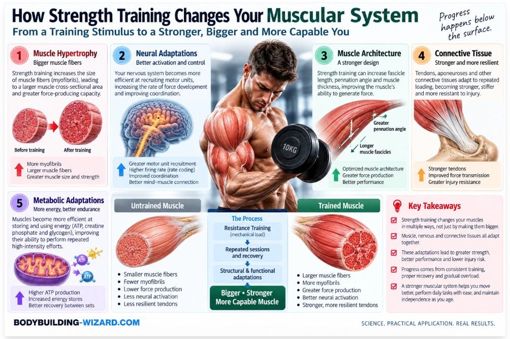
Muscle Hypertrophy
Muscle fibers increase in cross-sectional area, largely by adding contractile proteins (actin and myosin) inside existing fibers through ongoing exercise-driven protein synthesis and remodeling. We cover the full mechanism in Muscle Hypertrophy Explained: How Muscles Grow, and address the related question of whether fibers can also split and multiply in Muscle Hyperplasia: Can Humans Really Grow New Muscle Fibers?
Changes in Muscle Architecture
Training can increase pennation angle and fascicle length over time — architectural adaptations that differ depending on whether the training is concentric or eccentric in emphasis — which is part of how trained muscle becomes both bigger and better suited to force production.
Neural Adaptations
Especially early in a training program, much of the strength gain comes from improved motor unit recruitment, firing rate, and coordination — before significant hypertrophy has even occurred. This is why beginners often get dramatically stronger in the first weeks without visibly changing size.
Connective-Tissue Adaptations
Tendons and other connective tissue thicken and stiffen over time in response to consistent loading — a slower adaptation than muscle hypertrophy, which is one reason load should generally be progressed gradually. See Do Tendons Get Stronger? How Tendons Adapt to Strength Training for the full picture.
Metabolic Adaptations
Trained muscle becomes more efficient at storing and using energy substrates, and — depending on the training style — can improve either its capacity for sustained, oxidative work or its capacity for explosive, glycolytic work.
Two of the cellular mechanisms underlying long-term hypertrophy deserve their own dedicated pages: Satellite Cells Explained, and Muscle Nuclei (Myonuclei) Explained.
Muscle Growth vs. Strength: They Are Related, but Not the Same
Hypertrophy and strength correlate, but they aren’t identical. Strength also depends heavily on neural factors, technique, muscle architecture, and joint leverage — which is part of why two lifters with visually similar muscle size can have very different one-rep maxes, and why a bodybuilder’s training (higher volume, moderate loads, shorter rest) and a powerlifter’s training (lower volume, near-maximal loads, longer rest) both build muscle but optimize for different outcomes.
Why Anatomy Matters for Exercise Selection
Joint Action Matters More Than Exercise Names
An exercise’s name tells you almost nothing on its own — what matters is the joint action it produces. “Row” isn’t a muscle target; shoulder extension and scapular retraction are.
Muscle Function Helps Predict Which Exercises Train It
Once you know a muscle’s origin, insertion, and resulting joint action, you can predict — rather than guess — which exercises will meaningfully load it.
Anatomy Does Not Mean You Can Perfectly “Isolate” Every Muscle
Most exercises recruit multiple muscles simultaneously as agonists, synergists, and stabilizers, so true single-muscle isolation is more of a training ideal than a biomechanical reality.
Individual Anatomy Changes Exercise Execution
The same exercise can feel completely different from lifter to lifter — a lat pulldown, lateral raise, leg extension, Romanian deadlift, or curl will all be shaped by each individual’s limb length, joint structure, and attachment points.
Why Two Lifters May Feel the Same Exercise Differently
This is one of the most practically important — and most ignored — anatomical realities in the gym. Limb lengths, joint structure, tendon attachment points, mobility, technique, training history, and overall anthropometry all shape how a given exercise feels and which muscles it emphasizes for a specific person.
The takeaway: anatomy provides principles, not a single perfect exercise or technique that will work identically for everyone.
Coach’s Note: Anatomy Diagrams Are Not Exercise Prescriptions. In my years of coaching, one of the biggest mistakes I’ve seen lifters make is treating an anatomy diagram as if it were an exercise prescription. Knowing that a muscle performs a certain joint action is the starting point for exercise selection — not the end of it. I’ve had clients with nearly identical training histories respond completely differently to the same Romanian deadlift cue, simply because one had noticeably longer femurs relative to their torso. The anatomy told us why; it didn’t hand us a single “correct” technique for both of them.
Common Muscular System Myths
Myth: “Muscles push as well as pull.” Fact: skeletal muscles actively generate tensile (pulling) force rather than compressive (pushing) force — and that force generation isn’t limited to shortening. A muscle also generates tension isometrically, with no change in length, and eccentrically, while it lengthens. Apparent “pushing” movements happen because of how muscles are arranged around joints and levers, not because any muscle itself pushes.
Myth: “You can completely isolate one muscle.” Fact: as covered above, most exercises involve multiple agonists, synergists, and stabilizers working together — true isolation is rare.
Myth: “Soreness means a muscle was effectively trained.” Fact: delayed-onset muscle soreness is influenced heavily by novelty and eccentric loading, not simply by how effective a session was for hypertrophy or strength.
Myth: “More muscle always means proportionally more strength.” Fact: as covered in the “growth vs. strength” section above, neural factors, leverage, and technique mean size and strength don’t scale perfectly together.
Myth: “You need to feel a muscle working for it to be working.” Fact: mind-muscle connection can help in some contexts, but plenty of effective, heavily loaded compound lifts don’t produce an obvious “burn” in the target muscle while still driving real adaptation.
How to Apply Muscular System Anatomy to Your Training
This is the practical checklist version of everything above — the “Bodybuilder’s Take” of the entire article:
- Identify the joint action a lift is actually producing.
- Identify the primary muscles capable of producing that joint action.
- Consider the direction of resistance relative to that joint action throughout the range of motion.
- Consider muscle length and joint position at the point of greatest resistance.
- Account for your own individual anatomy rather than forcing a cue that works for someone else’s leverages.
- Progressively overload the movement over time.
- Judge exercises by the outcomes you’re getting, not by anatomy diagrams alone.
Key Takeaways
- ✓The muscular system includes skeletal, cardiac, and smooth muscle, but skeletal muscle is what lifters train and adapt.
- ✓A muscle is organized from whole muscle down to fascicles, fibers, myofibrils, sarcomeres, and actin/myosin filaments.
- ✓Force is transmitted through connective tissue (epimysium, perimysium, endomysium) into tendons and onto bone.
- ✓Contraction happens through the sliding filament mechanism, powered by ATP and triggered by calcium release.
- ✓No muscle works alone — agonists, antagonists, synergists, and stabilizers cooperate in virtually every movement.
- ✓Force output depends on more than muscle size — neural activation, joint angle, and leverage all matter.
- ✓Resistance training drives hypertrophy, neural adaptation, connective-tissue remodeling, and metabolic changes.
- ✓Anatomy gives you principles for exercise selection — not a single “correct” technique that fits every lifter.
Frequently Asked Questions
How many muscles are in the human body?
Most anatomy sources cite roughly 600 skeletal muscles, though the exact number varies depending on how certain muscles are classified or counted.
What are the three types of muscle tissue?
Skeletal muscle (voluntary, attached to bone), cardiac muscle (involuntary, found only in the heart), and smooth muscle (involuntary, found in organs and blood vessels).
What is the main function of the muscular system?
Its primary role is producing force and movement, but it also maintains posture, stabilizes joints, generates heat, supports circulation, and contributes to metabolic regulation.
What is the difference between a muscle and a muscle fiber?
A muscle (like the biceps brachii) is a whole organ made of thousands of individual muscle fibers, along with connective tissue, blood vessels, and nerves. A muscle fiber is a single, elongated muscle cell — one of the many building blocks that make up the whole muscle.
How do muscles make bones move?
A muscle contracts and shortens, pulling on the tendon connecting it to bone at its insertion point. Because bones act as levers around joints, this pull produces rotation or movement at that joint.
Do muscles push or only pull?
Muscles only generate tensile (pulling) force — they never actively push. This isn’t limited to shortening either: a muscle can generate this tension while shortening, while staying the same length (isometrically), or while lengthening (eccentrically). Movements that look like “pushing” happen because of how muscles are positioned relative to joints, not because any muscle is capable of pushing.
What happens to muscles when you lift weights?
In a single session, motor units are recruited, tension and energy demand increase, and metabolites accumulate, causing fatigue. Over weeks of consistent training, this drives hypertrophy, neural adaptations, connective-tissue remodeling, and metabolic changes.
Does strength training increase the number of muscle fibers?
In humans, most evidence indicates strength training primarily increases the size of existing muscle fibers (hypertrophy) rather than the number of fibers (hyperplasia). Whether meaningful fiber-number increases occur in humans remains debated and is covered in detail in our dedicated hyperplasia guide.
References
- Betts, J. G., et al. (2023). Anatomy and Physiology 2e. OpenStax. openstax.org/details/books/anatomy-and-physiology-2e
- Kenney, W. L., Wilmore, J. H., & Costill, D. L. (2021). Physiology of Sport and Exercise (8th ed.). Human Kinetics.
- Powers, S. K., & Howley, E. T. (2021). Exercise Physiology: Theory and Application to Fitness and Performance (11th ed.). McGraw Hill.
- Schoenfeld, B. J. (2010). The mechanisms of muscle hypertrophy and their application to resistance training. Journal of Strength and Conditioning Research. journals.lww.com/nsca-jscr
- Folland, J. P., & Williams, A. G. (2007). The adaptations to strength training: morphological and neurological contributions to increased strength. Sports Medicine. link.springer.com/article/10.2165/00007256-200737020-00004
- Lieber, R. L., & Fridén, J. (2000). Functional and clinical significance of skeletal muscle architecture. Muscle & Nerve. onlinelibrary.wiley.com
- Henneman, E., Somjen, G., & Carpenter, D. O. (1965). Functional significance of cell size in spinal motoneurons. Journal of Neurophysiology. journals.physiology.org/doi/10.1152/jn.1965.28.3.560
- Pedersen, B. K., & Febbraio, M. A. (2012). Muscles, exercise and obesity: skeletal muscle as a secretory organ. Nature Reviews Endocrinology. nature.com/articles/nrendo.2012.49
- Wackerhage, H., et al. (2019). Stimuli and sensors that initiate skeletal muscle hypertrophy following resistance exercise. Journal of Applied Physiology. journals.physiology.org
- American College of Sports Medicine. (2021). ACSM’s Guidelines for Exercise Testing and Prescription (11th ed.). Wolters Kluwer.
- Kjaer, M. (2004). Role of extracellular matrix in adaptation of tendon and skeletal muscle to mechanical loading. Physiological Reviews. journals.physiology.org
- Franchi, M. V., et al. (2014). Architectural, functional and molecular responses to concentric and eccentric loading in human skeletal muscle. Acta Physiologica. pubmed.ncbi.nlm.nih.gov/24387247
- Boyas, S., & Guével, A. (2011). Neuromuscular fatigue in healthy muscle: underlying factors and adaptation mechanisms. Annals of Physical and Rehabilitation Medicine. pubmed.ncbi.nlm.nih.gov/21376692
- Pedersen, B. K. (2013). Muscle as a secretory organ. Comprehensive Physiology. pubmed.ncbi.nlm.nih.gov/23897689
- McGlory, C., Devries, M. C., & Phillips, S. M. (2017). Skeletal muscle and resistance exercise training: the role of protein synthesis in recovery and remodeling. Journal of Applied Physiology. pubmed.ncbi.nlm.nih.gov/27742803
