
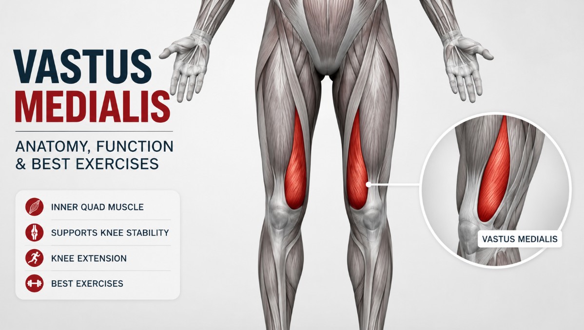
The “teardrop” muscle above the inner knee is the single most-referenced landmark of quad development in bodybuilding — but it’s also one of the most misunderstood, since most of what gets repeated about “activating the VMO” oversimplifies a genuinely two-part muscle with a real, if contested, clinical role in knee health.

Key Facts
The vastus medialis is the innermost of the four quadriceps muscles, forming the teardrop-shaped bulge above the inner knee. Its distal fibers — often called the vastus medialis oblique (VMO) — run at a steep angle and are largely responsible for pulling the patella medially, counterbalancing the pull of the vastus lateralis and helping keep the kneecap tracking properly during knee extension.
| Muscle type | One of four quadriceps femoris muscles |
|---|---|
| Primary function | Knee extension |
| Secondary function | Medial patellar stabilization (mainly via the oblique distal fibers, VMO) |
| Innervation | Femoral nerve (L2–L4) |
| Blood supply | Femoral artery, with contributions from the descending genicular artery |
| Common exercises | Leg extension (terminal range), front squat, leg press |
| Main bodybuilding role | The “teardrop” — a visible marker of complete quad development near the knee |
Vastus Medialis Anatomy & Location
The vastus medialis is one of four muscles that make up the quadriceps femoris group, alongside:
- Rectus femoris — the only quad head that crosses the hip as well as the knee
- Vastus lateralis — the largest and strongest of the four heads, forming the outer sweep
- Vastus intermedius — the deepest head, hidden entirely beneath the rectus femoris

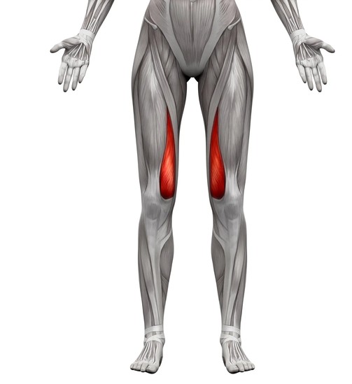
The vastus medialis runs along the inner thigh and becomes most visually distinct near the knee, where its lower fibers bulge into the recognizable teardrop shape lifters look for as a sign of complete quad development. To see exactly where it sits relative to the rest of your leg musculature, our interactive muscle anatomy tool lets you explore it alongside the other three heads.
Anatomically, the vastus medialis is usually described as having two regions rather than one: the vastus medialis longus (VML), whose fibers run nearly parallel to the femur, and the vastus medialis oblique (VMO), whose fibers run at a much steeper angle in the distal portion of the muscle. In roughly half of dissected specimens, there’s a visible fibro-fascial plane separating the two, though in practice they function as one continuous muscle.
Origin, Insertion, Innervation & Blood Supply
| Origin | Inferior intertrochanteric line, pectineal line, medial lip of the linea aspera, and medial supracondylar line of the femur; the most distal (VMO) fibers also arise from the adductor magnus tendon |
|---|---|
| Insertion | Quadriceps tendon, into the base and medial border of the patella, continuing via the patellar ligament to the tibial tuberosity; the distal oblique (VMO) fibers also blend into the medial patellar retinaculum |
| Innervation | Femoral nerve (L2–L4) |
| Blood supply | Femoral artery, with contributions from the descending genicular artery |
If terms like origin and insertion aren’t fully clear yet, check out our muscle anatomy basics guide for a plain-language breakdown of how muscles attach to bone and why it matters.
Vastus Medialis Structure & Muscle Architecture
The defining architectural feature of the vastus medialis is the sharp change in fiber angle between its two regions. Cadaveric studies have measured the vastus medialis longus fibers running at an average angle of only about 5 degrees relative to the shaft of the femur — nearly parallel, similar to the other vasti — while the vastus medialis oblique fibers run at a much steeper average angle of around 52 degrees. That difference in orientation is exactly what allows the VMO to pull the patella medially rather than simply adding to straight-line knee extension force.
This isn’t a minor technicality. Ultrasound studies comparing painful and pain-free knees have found the VMO’s cross-sectional area measurably smaller — by roughly 17-37%, depending on the measurement site — in patients with anterior knee pain, making VMO size a genuine, measurable marker used in patellofemoral pain research, not just a bodybuilding aesthetic term.
Functions of the Vastus Medialis
Like the vastus lateralis, the vastus medialis crosses only the knee joint, so its primary action is straightforward: knee extension, working alongside the other three quad heads. It doesn’t face the torque-sharing conflict that limits the rectus femoris in compound lifts, so it responds well to both compound and single-joint exercises.
Its secondary role, driven mainly by the oblique (VMO) fibers, is pulling the patella medially during the final degrees of knee extension — directly opposing the lateral pull of the vastus lateralis. This balance matters because the patella naturally tends to track laterally as the knee approaches full extension; a properly functioning VMO helps keep it centered in the femoral groove.
Vastus Medialis Synergists & Antagonists
Synergists (muscles that work alongside the vastus medialis to help produce the same movement):
Rectus femoris, vastus lateralis, and vastus intermedius — all three assist knee extension as fellow members of the quadriceps group.
Antagonists (muscles that oppose the vastus medialis’s action, working in the opposite direction):
The hamstrings oppose knee extension by flexing the knee. At the patella specifically, the vastus lateralis acts as a functional antagonist to the VMO, pulling the kneecap laterally rather than medially.
Common Vastus Medialis Exercises

| Exercise | Role of the Vastus Medialis |
|---|---|
| Leg extension (terminal range) | Strong contributor, particularly in the last 30 degrees of extension where research shows greater relative vastus medialis involvement |
| Front squat | Prime mover, alongside the other quad heads |
| Leg press | Prime mover, alongside the other quad heads |
| Terminal knee extension with external tibial rotation | Best-supported option for relatively biasing the vastus medialis over the vastus lateralis, per controlled EMG and cross-sectional research |
For a complete breakdown of quad training options, see our full guide: Best Quadriceps Exercises.
Bodybuilder’s Take
“Can I actually ‘activate’ my VMO with a special exercise, or is that a myth?” This is genuinely contested in the research. Older studies generally found no exercise selectively isolates the VMO from the rest of the quadriceps. However, more recent controlled research on terminal-range (final 30 degrees) knee extension performed with external leg rotation has found it can meaningfully improve the vastus medialis-to-vastus lateralis activity and cross-sectional ratio — so “VMO activation” isn’t pure myth, but it’s a narrower, more technical effect than the phrase usually implies in gym talk.
“Why do people say full range-of-motion squats build a better teardrop than partial reps?” The terminal-extension research above supports training through the final degrees of knee extension rather than stopping short, since that’s specifically where the vastus medialis, and especially the VMO, shows relatively greater involvement.
“My knee tracks oddly, and I’ve heard it’s a ‘weak VMO’ — is that really the cause?” VMO underdevelopment or delayed activation relative to the vastus lateralis is one documented factor associated with patellar maltracking and anterior knee pain, but it’s rarely the only cause — hip weakness, footwear, and overall training load also contribute. This is worth discussing with a physical therapist rather than self-diagnosing from gym folklore.
Common Vastus Medialis Conditions
This section is educational and should not replace professional medical advice.
Patellofemoral pain syndrome (PFPS) — a common cause of anterior knee pain, associated in the research literature with reduced VMO cross-sectional area and altered VMO-to-vastus lateralis activation timing, among other contributing factors.
Patellar maltracking — when the patella doesn’t glide smoothly within its groove during knee movement, sometimes linked to relative weakness or delayed firing of the VMO compared to the vastus lateralis.
Persistent anterior or medial knee pain, or a kneecap that feels unstable or “gives way,” should be evaluated by a healthcare professional rather than trained through.
Common Vastus Medialis Myths
Myth: You can never train the VMO differently from the rest of the quad. The classic view was that true selective activation is impossible, but newer controlled research on terminal-range knee extension with external rotation challenges that, showing a measurable shift in the vastus medialis-to-lateralis ratio.
Myth: A visible teardrop means your VMO is “strong” and injury-proof. Visual size is a reasonable proxy but not a guarantee — VMO function also depends on activation timing relative to the vastus lateralis, which isn’t something you can assess by looking in a mirror.
Complete your anatomy knowledge with the muscles of the chest, shoulders, back, arms, core, and neck.
Explore Upper Body MusclesKey Takeaways
- The vastus medialis has two functional regions — the VML (nearly parallel fibers) and the VMO (steeply angled fibers) — that behave differently.
- The VMO’s main job is pulling the patella medially, counterbalancing the lateral pull of the vastus lateralis.
- Reduced VMO size is a measurable factor associated with patellofemoral pain in research, not just a bodybuilding aesthetic concern.
- True VMO isolation was long considered impossible, but newer research on terminal-range knee extension with external rotation shows a measurable relative shift is achievable.
- Training through full range of motion, especially the final degrees of knee extension, matters more for this muscle than for the other quad heads.
Frequently Asked Questions
What is the VMO?
The vastus medialis oblique — the distal, steeply angled fibers of the vastus medialis that are primarily responsible for pulling the patella medially during knee extension.
Can you isolate the VMO with a specific exercise?
Not completely, but recent research on terminal-range knee extension performed with external leg rotation has shown a measurable relative shift toward greater vastus medialis involvement.
Why is the vastus medialis called the “teardrop” muscle?
Its lower fibers form a visible teardrop-shaped bulge above the inner knee when well developed, which lifters commonly use as a visual marker of complete quad development.
Is a weak VMO the cause of knee pain?
It can be a contributing factor in patellofemoral pain and patellar maltracking, but it’s rarely the sole cause — hip strength, footwear, and overall training load also play a role.
Does full range of motion matter more for the vastus medialis than for other quad muscles?
Research on terminal knee extension suggests the vastus medialis, and especially the VMO, shows relatively greater involvement in the final degrees of extension, which supports training through a full range rather than stopping short.
Sources & References
Every claim in this guide is grounded in anatomical references and published research.
